Potassium »
PDB 7olt-7pji »
7on2 »
Potassium in PDB 7on2: Saftsz Complexed with Gdp (Soaking in 10 Mm Cydta)
Protein crystallography data
The structure of Saftsz Complexed with Gdp (Soaking in 10 Mm Cydta), PDB code: 7on2
was solved by
C.Fernandez-Tornero,
F.M.Ruiz,
J.M.Andreu,
with X-Ray Crystallography technique. A brief refinement statistics is given in the table below:
| Resolution Low / High (Å) | 40.98 / 1.69 |
| Space group | C 1 2 1 |
| Cell size a, b, c (Å), α, β, γ (°) | 72.402, 50.207, 88.05, 90, 111.42, 90 |
| R / Rfree (%) | 20.4 / 24.7 |
Potassium Binding Sites:
The binding sites of Potassium atom in the Saftsz Complexed with Gdp (Soaking in 10 Mm Cydta)
(pdb code 7on2). This binding sites where shown within
5.0 Angstroms radius around Potassium atom.
In total only one binding site of Potassium was determined in the Saftsz Complexed with Gdp (Soaking in 10 Mm Cydta), PDB code: 7on2:
In total only one binding site of Potassium was determined in the Saftsz Complexed with Gdp (Soaking in 10 Mm Cydta), PDB code: 7on2:
Potassium binding site 1 out of 1 in 7on2
Go back to
Potassium binding site 1 out
of 1 in the Saftsz Complexed with Gdp (Soaking in 10 Mm Cydta)
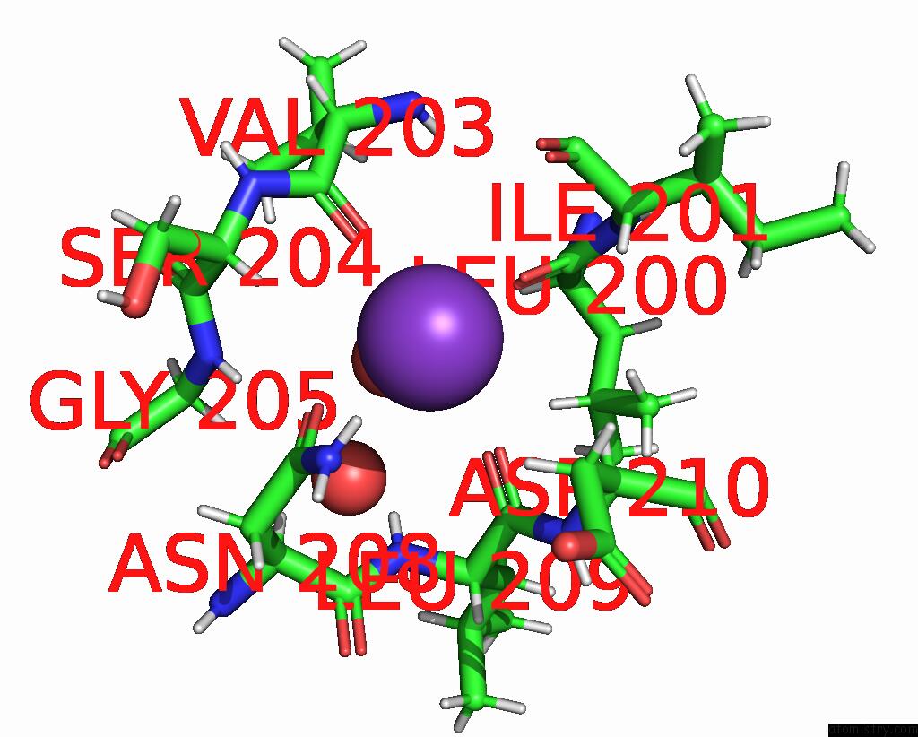
Mono view
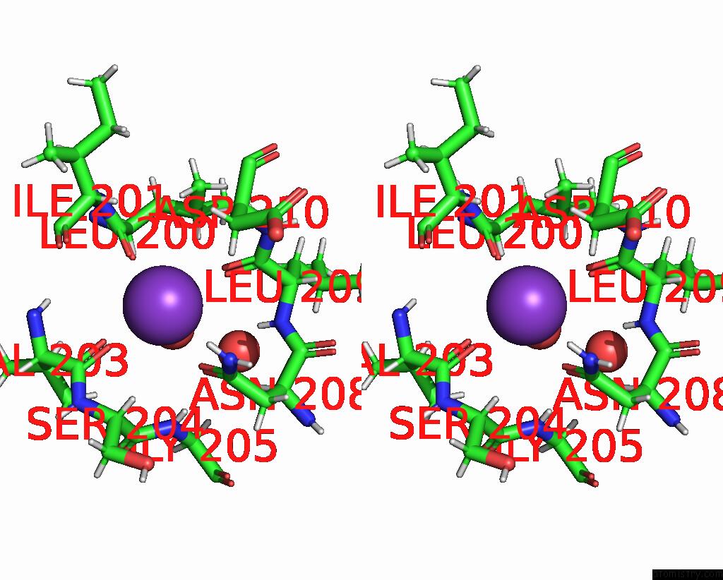
Stereo pair view
Mono view
Stereo pair view
A full contact list of Potassium with other atoms in the K binding
site number 1 of Saftsz Complexed with Gdp (Soaking in 10 Mm Cydta) within 5.0Å range:
|
Reference:
F.M.Ruiz,
S.Huecas,
A.Santos-Aledo,
E.A.Prim,
J.M.Andreu,
C.Fernandez-Tornero.
Ftsz Filament Structures in Different Nucleotide States Reveal the Mechanism of Assembly Dynamics. Plos Biol. V. 20 01497 2022.
ISSN: ESSN 1545-7885
PubMed: 35312677
DOI: 10.1371/JOURNAL.PBIO.3001497
Page generated: Sat Aug 9 13:52:06 2025
ISSN: ESSN 1545-7885
PubMed: 35312677
DOI: 10.1371/JOURNAL.PBIO.3001497
Last articles
Mg in 2PS4Mg in 2PRC
Mg in 2PS1
Mg in 2PRY
Mg in 2PRN
Mg in 2PPQ
Mg in 2PPB
Mg in 2PLY
Mg in 2PNQ
Mg in 2PP3