Potassium »
PDB 7k5h-7lo2 »
7kz0 »
Potassium in PDB 7kz0: Human MBD4 Glycosylase Domain Bound to Dna Containing Substrate Analog 2'-Deoxy-Pseudouridine
Protein crystallography data
The structure of Human MBD4 Glycosylase Domain Bound to Dna Containing Substrate Analog 2'-Deoxy-Pseudouridine, PDB code: 7kz0
was solved by
L.S.Pidugu,
E.Pozharski,
A.C.Drohat,
with X-Ray Crystallography technique. A brief refinement statistics is given in the table below:
| Resolution Low / High (Å) | 38.65 / 1.57 |
| Space group | P 21 21 21 |
| Cell size a, b, c (Å), α, β, γ (°) | 41.361, 56.86, 105.416, 90, 90, 90 |
| R / Rfree (%) | 18.8 / 21.5 |
Potassium Binding Sites:
The binding sites of Potassium atom in the Human MBD4 Glycosylase Domain Bound to Dna Containing Substrate Analog 2'-Deoxy-Pseudouridine
(pdb code 7kz0). This binding sites where shown within
5.0 Angstroms radius around Potassium atom.
In total only one binding site of Potassium was determined in the Human MBD4 Glycosylase Domain Bound to Dna Containing Substrate Analog 2'-Deoxy-Pseudouridine, PDB code: 7kz0:
In total only one binding site of Potassium was determined in the Human MBD4 Glycosylase Domain Bound to Dna Containing Substrate Analog 2'-Deoxy-Pseudouridine, PDB code: 7kz0:
Potassium binding site 1 out of 1 in 7kz0
Go back to
Potassium binding site 1 out
of 1 in the Human MBD4 Glycosylase Domain Bound to Dna Containing Substrate Analog 2'-Deoxy-Pseudouridine
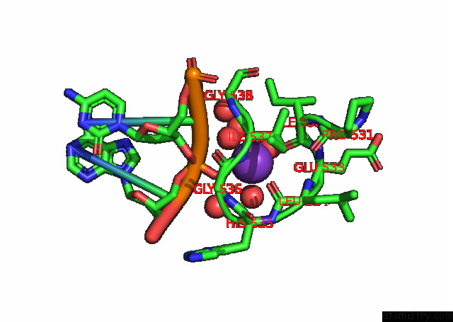
Mono view
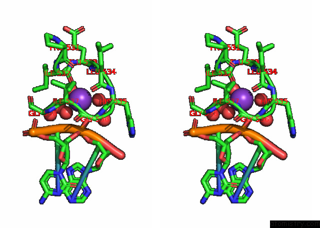
Stereo pair view
Mono view
Stereo pair view
A full contact list of Potassium with other atoms in the K binding
site number 1 of Human MBD4 Glycosylase Domain Bound to Dna Containing Substrate Analog 2'-Deoxy-Pseudouridine within 5.0Å range:
|
Reference:
L.S.Pidugu,
H.Bright,
W.J.Lin,
C.Majumdar,
R.P.Van Ostrand,
S.S.David,
E.Pozharski,
A.C.Drohat.
Structural Insights Into the Mechanism of Base Excision By MBD4. J.Mol.Biol. V. 433 67097 2021.
ISSN: ESSN 1089-8638
PubMed: 34107280
DOI: 10.1016/J.JMB.2021.167097
Page generated: Mon Aug 12 19:18:56 2024
ISSN: ESSN 1089-8638
PubMed: 34107280
DOI: 10.1016/J.JMB.2021.167097
Last articles
Zn in 9JYWZn in 9IR4
Zn in 9IR3
Zn in 9GMX
Zn in 9GMW
Zn in 9JEJ
Zn in 9ERF
Zn in 9ERE
Zn in 9EGV
Zn in 9EGW