Potassium »
PDB 7jvv-7kw7 »
7kd9 »
Potassium in PDB 7kd9: Crystal Structure of Gallic Acid Decarboxylase From Arxula Adeninivorans
Protein crystallography data
The structure of Crystal Structure of Gallic Acid Decarboxylase From Arxula Adeninivorans, PDB code: 7kd9
was solved by
M.Zeug,
N.Markovic,
C.V.Iancu,
J.Tripp,
M.Oreb,
J.Choe,
with X-Ray Crystallography technique. A brief refinement statistics is given in the table below:
| Resolution Low / High (Å) | 46.61 / 1.94 |
| Space group | P 21 21 2 |
| Cell size a, b, c (Å), α, β, γ (°) | 91.632, 265.301, 93.728, 90, 90, 90 |
| R / Rfree (%) | 19.1 / 23.8 |
Potassium Binding Sites:
The binding sites of Potassium atom in the Crystal Structure of Gallic Acid Decarboxylase From Arxula Adeninivorans
(pdb code 7kd9). This binding sites where shown within
5.0 Angstroms radius around Potassium atom.
In total 3 binding sites of Potassium where determined in the Crystal Structure of Gallic Acid Decarboxylase From Arxula Adeninivorans, PDB code: 7kd9:
Jump to Potassium binding site number: 1; 2; 3;
In total 3 binding sites of Potassium where determined in the Crystal Structure of Gallic Acid Decarboxylase From Arxula Adeninivorans, PDB code: 7kd9:
Jump to Potassium binding site number: 1; 2; 3;
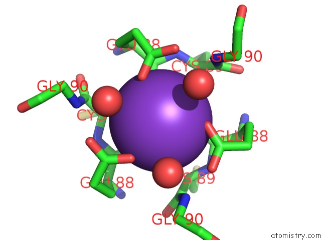
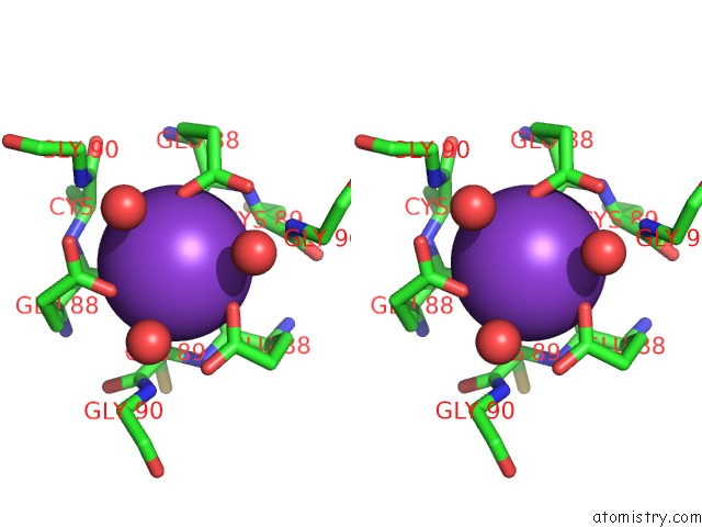
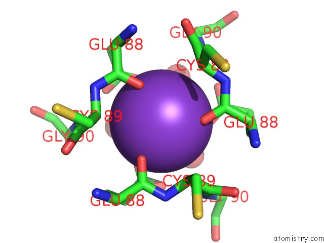
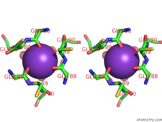
Potassium binding site 1 out of 3 in 7kd9
Go back to
Potassium binding site 1 out
of 3 in the Crystal Structure of Gallic Acid Decarboxylase From Arxula Adeninivorans
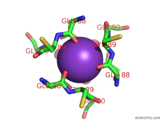
Mono view
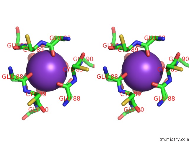
Stereo pair view
Mono view
Stereo pair view
A full contact list of Potassium with other atoms in the K binding
site number 1 of Crystal Structure of Gallic Acid Decarboxylase From Arxula Adeninivorans within 5.0Å range:
|
Potassium binding site 2 out of 3 in 7kd9
Go back to
Potassium binding site 2 out
of 3 in the Crystal Structure of Gallic Acid Decarboxylase From Arxula Adeninivorans
Mono view
Stereo pair view
Mono view
Stereo pair view
A full contact list of Potassium with other atoms in the K binding
site number 2 of Crystal Structure of Gallic Acid Decarboxylase From Arxula Adeninivorans within 5.0Å range:
|
Potassium binding site 3 out of 3 in 7kd9
Go back to
Potassium binding site 3 out
of 3 in the Crystal Structure of Gallic Acid Decarboxylase From Arxula Adeninivorans
Mono view
Stereo pair view
Mono view
Stereo pair view
A full contact list of Potassium with other atoms in the K binding
site number 3 of Crystal Structure of Gallic Acid Decarboxylase From Arxula Adeninivorans within 5.0Å range:
|
Reference:
M.Zeug,
N.Markovic,
C.V.Iancu,
J.Tripp,
M.Oreb,
J.Y.Choe.
Crystal Structures of Non-Oxidative Decarboxylases Reveal A New Mechanism of Action with A Catalytic Dyad and Structural Twists. Sci Rep V. 11 3056 2021.
ISSN: ESSN 2045-2322
PubMed: 33542397
DOI: 10.1038/S41598-021-82660-Z
Page generated: Sat Aug 9 13:31:22 2025
ISSN: ESSN 2045-2322
PubMed: 33542397
DOI: 10.1038/S41598-021-82660-Z
Last articles
Mg in 5G0RMg in 5G5V
Mg in 5G3T
Mg in 5G5T
Mg in 5G5S
Mg in 5G4A
Mg in 5G57
Mg in 5G50
Mg in 5G41
Mg in 5G3Z