Potassium »
PDB 7akk-7byn »
7aoa »
Potassium in PDB 7aoa: Structure of the Extended MTA1/HDAC1/MBD2/RBBP4 Nurd Deacetylase Complex
Enzymatic activity of Structure of the Extended MTA1/HDAC1/MBD2/RBBP4 Nurd Deacetylase Complex
All present enzymatic activity of Structure of the Extended MTA1/HDAC1/MBD2/RBBP4 Nurd Deacetylase Complex:
3.5.1.98;
3.5.1.98;
Other elements in 7aoa:
The structure of Structure of the Extended MTA1/HDAC1/MBD2/RBBP4 Nurd Deacetylase Complex also contains other interesting chemical elements:
| Zinc | (Zn) | 2 atoms |
Potassium Binding Sites:
The binding sites of Potassium atom in the Structure of the Extended MTA1/HDAC1/MBD2/RBBP4 Nurd Deacetylase Complex
(pdb code 7aoa). This binding sites where shown within
5.0 Angstroms radius around Potassium atom.
In total 4 binding sites of Potassium where determined in the Structure of the Extended MTA1/HDAC1/MBD2/RBBP4 Nurd Deacetylase Complex, PDB code: 7aoa:
Jump to Potassium binding site number: 1; 2; 3; 4;
In total 4 binding sites of Potassium where determined in the Structure of the Extended MTA1/HDAC1/MBD2/RBBP4 Nurd Deacetylase Complex, PDB code: 7aoa:
Jump to Potassium binding site number: 1; 2; 3; 4;
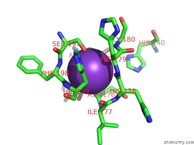
Potassium binding site 1 out of 4 in 7aoa
Go back to
Potassium binding site 1 out
of 4 in the Structure of the Extended MTA1/HDAC1/MBD2/RBBP4 Nurd Deacetylase Complex
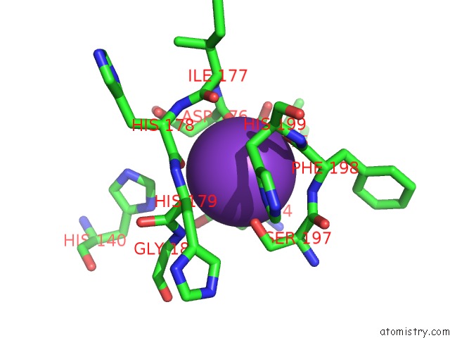
Mono view
Stereo pair view
Mono view
Stereo pair view
A full contact list of Potassium with other atoms in the K binding
site number 1 of Structure of the Extended MTA1/HDAC1/MBD2/RBBP4 Nurd Deacetylase Complex within 5.0Å range:
|
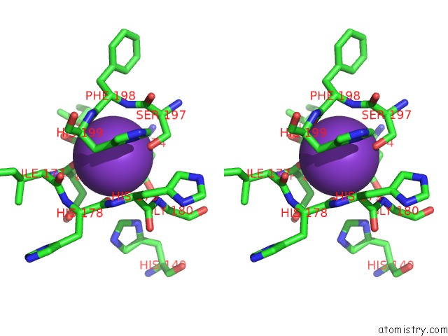
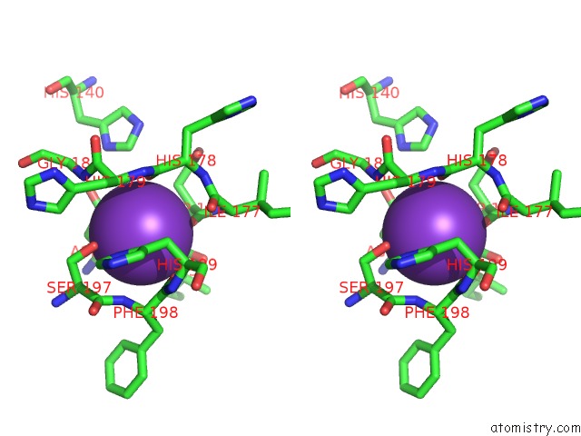
Potassium binding site 2 out of 4 in 7aoa
Go back to
Potassium binding site 2 out
of 4 in the Structure of the Extended MTA1/HDAC1/MBD2/RBBP4 Nurd Deacetylase Complex
Mono view
Stereo pair view
Mono view
Stereo pair view
A full contact list of Potassium with other atoms in the K binding
site number 2 of Structure of the Extended MTA1/HDAC1/MBD2/RBBP4 Nurd Deacetylase Complex within 5.0Å range:
|
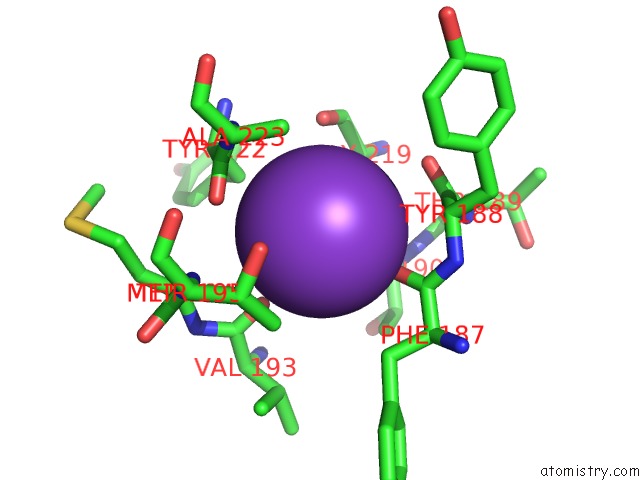
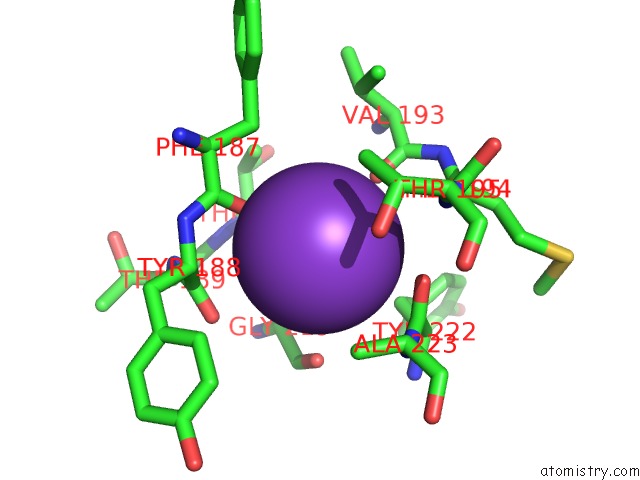
Potassium binding site 3 out of 4 in 7aoa
Go back to
Potassium binding site 3 out
of 4 in the Structure of the Extended MTA1/HDAC1/MBD2/RBBP4 Nurd Deacetylase Complex
Mono view
Stereo pair view
Mono view
Stereo pair view
A full contact list of Potassium with other atoms in the K binding
site number 3 of Structure of the Extended MTA1/HDAC1/MBD2/RBBP4 Nurd Deacetylase Complex within 5.0Å range:
|
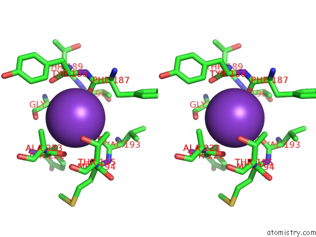
Potassium binding site 4 out of 4 in 7aoa
Go back to
Potassium binding site 4 out
of 4 in the Structure of the Extended MTA1/HDAC1/MBD2/RBBP4 Nurd Deacetylase Complex
Mono view
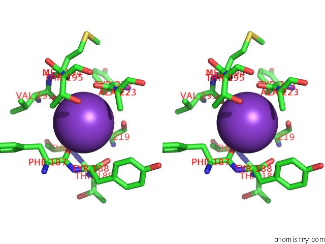
Stereo pair view
Mono view
Stereo pair view
A full contact list of Potassium with other atoms in the K binding
site number 4 of Structure of the Extended MTA1/HDAC1/MBD2/RBBP4 Nurd Deacetylase Complex within 5.0Å range:
|
Reference:
C.J.Millard,
L.Fairall,
T.J.Ragan,
C.G.Savva,
J.W.R.Schwabe.
The Topology of Chromatin-Binding Domains in the Nurd Deacetylase Complex To Be Published.
Page generated: Sat Aug 9 12:58:21 2025
Last articles
Mg in 1YM0Mg in 1YLS
Mg in 1YL7
Mg in 1YL6
Mg in 1YL5
Mg in 1YKV
Mg in 1YKQ
Mg in 1YIT
Mg in 1YIJ
Mg in 1YJF