Potassium »
PDB 6z7v-7adi »
6z8x »
Potassium in PDB 6z8x: X-Ray Structure of the Complex Between Human Alpha Thrombin and A Thrombin Binding Aptamer Variant (Tba-3LEU), Which Contains Leucyl Amide in the Side Chain of THY3 at N3.
Enzymatic activity of X-Ray Structure of the Complex Between Human Alpha Thrombin and A Thrombin Binding Aptamer Variant (Tba-3LEU), Which Contains Leucyl Amide in the Side Chain of THY3 at N3.
All present enzymatic activity of X-Ray Structure of the Complex Between Human Alpha Thrombin and A Thrombin Binding Aptamer Variant (Tba-3LEU), Which Contains Leucyl Amide in the Side Chain of THY3 at N3.:
3.4.21.5;
3.4.21.5;
Protein crystallography data
The structure of X-Ray Structure of the Complex Between Human Alpha Thrombin and A Thrombin Binding Aptamer Variant (Tba-3LEU), Which Contains Leucyl Amide in the Side Chain of THY3 at N3., PDB code: 6z8x
was solved by
R.Troisi,
E.N.Timofeev,
F.Sica,
with X-Ray Crystallography technique. A brief refinement statistics is given in the table below:
| Resolution Low / High (Å) | 81.99 / 2.53 |
| Space group | P 32 2 1 |
| Cell size a, b, c (Å), α, β, γ (°) | 94.671, 94.671, 124.693, 90, 90, 120 |
| R / Rfree (%) | 24.5 / 29.2 |
Other elements in 6z8x:
The structure of X-Ray Structure of the Complex Between Human Alpha Thrombin and A Thrombin Binding Aptamer Variant (Tba-3LEU), Which Contains Leucyl Amide in the Side Chain of THY3 at N3. also contains other interesting chemical elements:
| Sodium | (Na) | 1 atom |
Potassium Binding Sites:
The binding sites of Potassium atom in the X-Ray Structure of the Complex Between Human Alpha Thrombin and A Thrombin Binding Aptamer Variant (Tba-3LEU), Which Contains Leucyl Amide in the Side Chain of THY3 at N3.
(pdb code 6z8x). This binding sites where shown within
5.0 Angstroms radius around Potassium atom.
In total only one binding site of Potassium was determined in the X-Ray Structure of the Complex Between Human Alpha Thrombin and A Thrombin Binding Aptamer Variant (Tba-3LEU), Which Contains Leucyl Amide in the Side Chain of THY3 at N3., PDB code: 6z8x:
In total only one binding site of Potassium was determined in the X-Ray Structure of the Complex Between Human Alpha Thrombin and A Thrombin Binding Aptamer Variant (Tba-3LEU), Which Contains Leucyl Amide in the Side Chain of THY3 at N3., PDB code: 6z8x:
Potassium binding site 1 out of 1 in 6z8x
Go back to
Potassium binding site 1 out
of 1 in the X-Ray Structure of the Complex Between Human Alpha Thrombin and A Thrombin Binding Aptamer Variant (Tba-3LEU), Which Contains Leucyl Amide in the Side Chain of THY3 at N3.
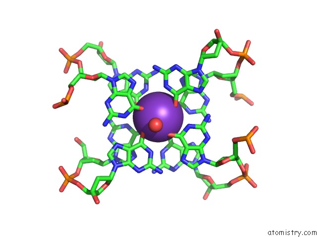
Mono view
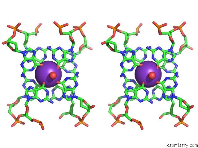
Stereo pair view
Mono view
Stereo pair view
A full contact list of Potassium with other atoms in the K binding
site number 1 of X-Ray Structure of the Complex Between Human Alpha Thrombin and A Thrombin Binding Aptamer Variant (Tba-3LEU), Which Contains Leucyl Amide in the Side Chain of THY3 at N3. within 5.0Å range:
|
Reference:
I.Smirnov,
N.Kolganova,
R.Troisi,
F.Sica,
E.Timofeev.
Expanding the Recognition Interface of the Thrombin Binding Aptamer HD1 Through Modification of Residues T3 and T12 Mol.Ther. 2021.
ISSN: ESSN 1525-0024
DOI: 10.1016/J.OMTN.2021.01.004
Page generated: Mon Aug 12 18:31:27 2024
ISSN: ESSN 1525-0024
DOI: 10.1016/J.OMTN.2021.01.004
Last articles
Zn in 9J0NZn in 9J0O
Zn in 9J0P
Zn in 9FJX
Zn in 9EKB
Zn in 9C0F
Zn in 9CAH
Zn in 9CH0
Zn in 9CH3
Zn in 9CH1