Potassium »
PDB 6w87-6x1j »
6wbq »
Potassium in PDB 6wbq: Crystal Structure of Danio Rerio Histone Deacetylase 10 in Complex with Tubastatin A
Enzymatic activity of Crystal Structure of Danio Rerio Histone Deacetylase 10 in Complex with Tubastatin A
All present enzymatic activity of Crystal Structure of Danio Rerio Histone Deacetylase 10 in Complex with Tubastatin A:
3.5.1.48; 3.5.1.62;
3.5.1.48; 3.5.1.62;
Protein crystallography data
The structure of Crystal Structure of Danio Rerio Histone Deacetylase 10 in Complex with Tubastatin A, PDB code: 6wbq
was solved by
C.J.Herbst-Gervasoni,
D.W.Christianson,
with X-Ray Crystallography technique. A brief refinement statistics is given in the table below:
| Resolution Low / High (Å) | 53.24 / 2.00 |
| Space group | P 31 2 1 |
| Cell size a, b, c (Å), α, β, γ (°) | 80.630, 80.630, 246.811, 90.00, 90.00, 120.00 |
| R / Rfree (%) | 19.9 / 23.5 |
Other elements in 6wbq:
The structure of Crystal Structure of Danio Rerio Histone Deacetylase 10 in Complex with Tubastatin A also contains other interesting chemical elements:
| Zinc | (Zn) | 1 atom |
Potassium Binding Sites:
The binding sites of Potassium atom in the Crystal Structure of Danio Rerio Histone Deacetylase 10 in Complex with Tubastatin A
(pdb code 6wbq). This binding sites where shown within
5.0 Angstroms radius around Potassium atom.
In total 2 binding sites of Potassium where determined in the Crystal Structure of Danio Rerio Histone Deacetylase 10 in Complex with Tubastatin A, PDB code: 6wbq:
Jump to Potassium binding site number: 1; 2;
In total 2 binding sites of Potassium where determined in the Crystal Structure of Danio Rerio Histone Deacetylase 10 in Complex with Tubastatin A, PDB code: 6wbq:
Jump to Potassium binding site number: 1; 2;
Potassium binding site 1 out of 2 in 6wbq
Go back to
Potassium binding site 1 out
of 2 in the Crystal Structure of Danio Rerio Histone Deacetylase 10 in Complex with Tubastatin A
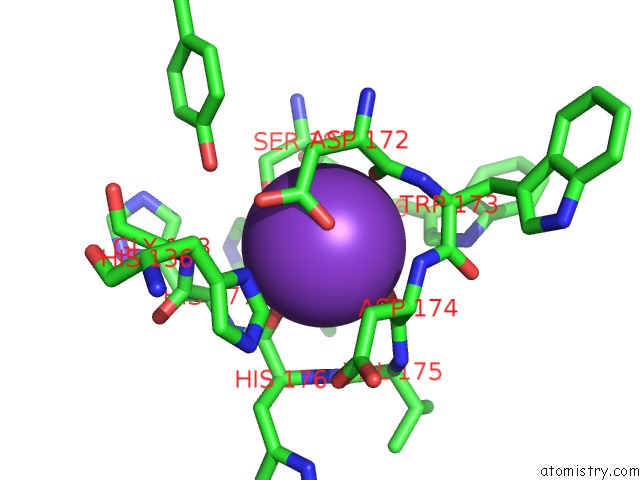
Mono view
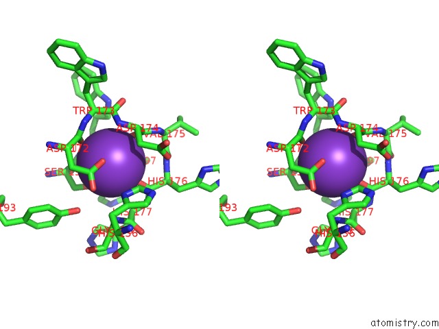
Stereo pair view
Mono view
Stereo pair view
A full contact list of Potassium with other atoms in the K binding
site number 1 of Crystal Structure of Danio Rerio Histone Deacetylase 10 in Complex with Tubastatin A within 5.0Å range:
|
Potassium binding site 2 out of 2 in 6wbq
Go back to
Potassium binding site 2 out
of 2 in the Crystal Structure of Danio Rerio Histone Deacetylase 10 in Complex with Tubastatin A
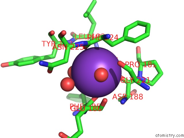
Mono view
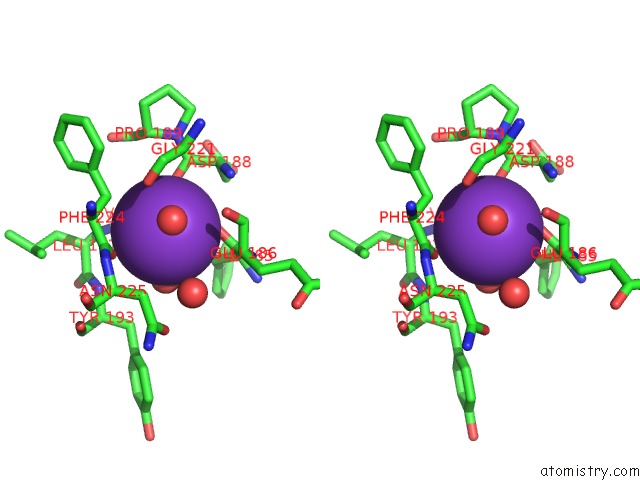
Stereo pair view
Mono view
Stereo pair view
A full contact list of Potassium with other atoms in the K binding
site number 2 of Crystal Structure of Danio Rerio Histone Deacetylase 10 in Complex with Tubastatin A within 5.0Å range:
|
Reference:
C.J.Herbst-Gervasoni,
R.R.Steimbach,
M.Morgen,
A.K.Miller,
D.W.Christianson.
Structural Basis For the Selective Inhibition of HDAC10, the Cytosolic Polyamine Deacetylase. Acs Chem.Biol. V. 15 2154 2020.
ISSN: ESSN 1554-8937
PubMed: 32659072
DOI: 10.1021/ACSCHEMBIO.0C00362
Page generated: Sat Aug 9 12:36:55 2025
ISSN: ESSN 1554-8937
PubMed: 32659072
DOI: 10.1021/ACSCHEMBIO.0C00362
Last articles
Mg in 2HNYMg in 2HMF
Mg in 2HMC
Mg in 2HND
Mg in 2HMA
Mg in 2HJ6
Mg in 2HK6
Mg in 2HKJ
Mg in 2HIT
Mg in 2HJP