Potassium »
PDB 6v8y-6w86 »
6vua »
Potassium in PDB 6vua: X-Ray Structure of Human CD38 Catalytic Domain with 2'-Cl-Aranad+
Enzymatic activity of X-Ray Structure of Human CD38 Catalytic Domain with 2'-Cl-Aranad+
All present enzymatic activity of X-Ray Structure of Human CD38 Catalytic Domain with 2'-Cl-Aranad+:
2.4.99.20; 3.2.2.6;
2.4.99.20; 3.2.2.6;
Protein crystallography data
The structure of X-Ray Structure of Human CD38 Catalytic Domain with 2'-Cl-Aranad+, PDB code: 6vua
was solved by
Z.Dai,
X.N.Zhang,
F.Nasertorabi,
G.W.Han,
R.C.Stevens,
Y.Zhang,
with X-Ray Crystallography technique. A brief refinement statistics is given in the table below:
| Resolution Low / High (Å) | 28.71 / 1.50 |
| Space group | P 41 2 2 |
| Cell size a, b, c (Å), α, β, γ (°) | 114.762, 114.762, 97.154, 90.00, 90.00, 90.00 |
| R / Rfree (%) | 16.6 / 19 |
Other elements in 6vua:
The structure of X-Ray Structure of Human CD38 Catalytic Domain with 2'-Cl-Aranad+ also contains other interesting chemical elements:
| Chlorine | (Cl) | 2 atoms |
Potassium Binding Sites:
The binding sites of Potassium atom in the X-Ray Structure of Human CD38 Catalytic Domain with 2'-Cl-Aranad+
(pdb code 6vua). This binding sites where shown within
5.0 Angstroms radius around Potassium atom.
In total 2 binding sites of Potassium where determined in the X-Ray Structure of Human CD38 Catalytic Domain with 2'-Cl-Aranad+, PDB code: 6vua:
Jump to Potassium binding site number: 1; 2;
In total 2 binding sites of Potassium where determined in the X-Ray Structure of Human CD38 Catalytic Domain with 2'-Cl-Aranad+, PDB code: 6vua:
Jump to Potassium binding site number: 1; 2;
Potassium binding site 1 out of 2 in 6vua
Go back to
Potassium binding site 1 out
of 2 in the X-Ray Structure of Human CD38 Catalytic Domain with 2'-Cl-Aranad+
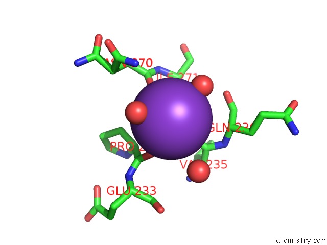
Mono view
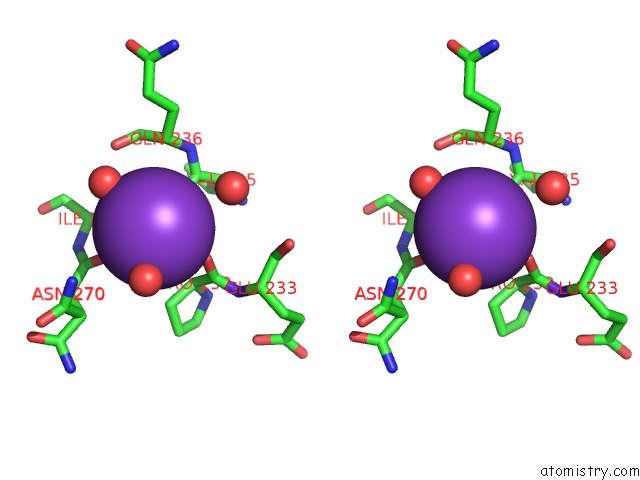
Stereo pair view
Mono view
Stereo pair view
A full contact list of Potassium with other atoms in the K binding
site number 1 of X-Ray Structure of Human CD38 Catalytic Domain with 2'-Cl-Aranad+ within 5.0Å range:
|
Potassium binding site 2 out of 2 in 6vua
Go back to
Potassium binding site 2 out
of 2 in the X-Ray Structure of Human CD38 Catalytic Domain with 2'-Cl-Aranad+
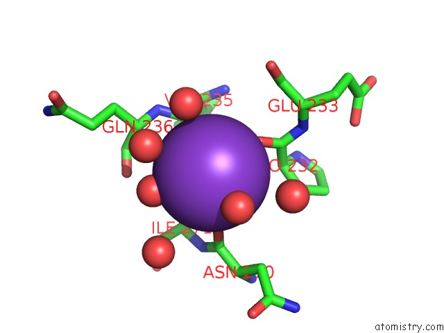
Mono view
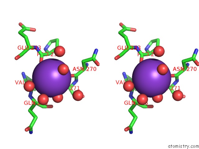
Stereo pair view
Mono view
Stereo pair view
A full contact list of Potassium with other atoms in the K binding
site number 2 of X-Ray Structure of Human CD38 Catalytic Domain with 2'-Cl-Aranad+ within 5.0Å range:
|
Reference:
Z.Dai,
X.N.Zhang,
F.Nasertorabi,
Q.Cheng,
J.Li,
B.B.Katz,
G.Smbatyan,
H.Pei,
S.G.Louie,
H.-J.Lenz,
R.C.Stevens,
Y.Zhang.
Synthesis of Site-Specific Antibody-Drug Conjugates By Adp-Ribosyl Cyclases To Be Published.
Page generated: Mon Aug 12 18:01:29 2024
Last articles
Zn in 9J0NZn in 9J0O
Zn in 9J0P
Zn in 9FJX
Zn in 9EKB
Zn in 9C0F
Zn in 9CAH
Zn in 9CH0
Zn in 9CH3
Zn in 9CH1