Potassium »
PDB 6v8y-6w86 »
6vnq »
Potassium in PDB 6vnq: Crystal Structure of Danio Rerio Histone Deacetylase 10 in Complex with Bishydroxamic Acid Based Inhibitor
Enzymatic activity of Crystal Structure of Danio Rerio Histone Deacetylase 10 in Complex with Bishydroxamic Acid Based Inhibitor
All present enzymatic activity of Crystal Structure of Danio Rerio Histone Deacetylase 10 in Complex with Bishydroxamic Acid Based Inhibitor:
3.5.1.48; 3.5.1.62;
3.5.1.48; 3.5.1.62;
Protein crystallography data
The structure of Crystal Structure of Danio Rerio Histone Deacetylase 10 in Complex with Bishydroxamic Acid Based Inhibitor, PDB code: 6vnq
was solved by
C.J.Herbst-Gervasoni,
D.W.Christianson,
with X-Ray Crystallography technique. A brief refinement statistics is given in the table below:
| Resolution Low / High (Å) | 69.76 / 2.05 |
| Space group | P 31 2 1 |
| Cell size a, b, c (Å), α, β, γ (°) | 80.549, 80.549, 245.370, 90.00, 90.00, 120.00 |
| R / Rfree (%) | 19.3 / 23.1 |
Other elements in 6vnq:
The structure of Crystal Structure of Danio Rerio Histone Deacetylase 10 in Complex with Bishydroxamic Acid Based Inhibitor also contains other interesting chemical elements:
| Zinc | (Zn) | 1 atom |
Potassium Binding Sites:
The binding sites of Potassium atom in the Crystal Structure of Danio Rerio Histone Deacetylase 10 in Complex with Bishydroxamic Acid Based Inhibitor
(pdb code 6vnq). This binding sites where shown within
5.0 Angstroms radius around Potassium atom.
In total 2 binding sites of Potassium where determined in the Crystal Structure of Danio Rerio Histone Deacetylase 10 in Complex with Bishydroxamic Acid Based Inhibitor, PDB code: 6vnq:
Jump to Potassium binding site number: 1; 2;
In total 2 binding sites of Potassium where determined in the Crystal Structure of Danio Rerio Histone Deacetylase 10 in Complex with Bishydroxamic Acid Based Inhibitor, PDB code: 6vnq:
Jump to Potassium binding site number: 1; 2;
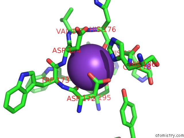
Potassium binding site 1 out of 2 in 6vnq
Go back to
Potassium binding site 1 out
of 2 in the Crystal Structure of Danio Rerio Histone Deacetylase 10 in Complex with Bishydroxamic Acid Based Inhibitor
Mono view
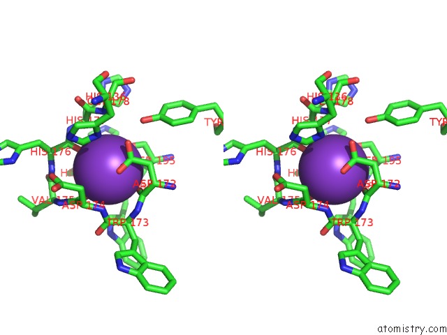
Stereo pair view
Mono view
Stereo pair view
A full contact list of Potassium with other atoms in the K binding
site number 1 of Crystal Structure of Danio Rerio Histone Deacetylase 10 in Complex with Bishydroxamic Acid Based Inhibitor within 5.0Å range:
|
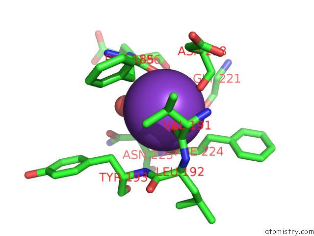
Potassium binding site 2 out of 2 in 6vnq
Go back to
Potassium binding site 2 out
of 2 in the Crystal Structure of Danio Rerio Histone Deacetylase 10 in Complex with Bishydroxamic Acid Based Inhibitor
Mono view
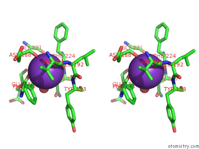
Stereo pair view
Mono view
Stereo pair view
A full contact list of Potassium with other atoms in the K binding
site number 2 of Crystal Structure of Danio Rerio Histone Deacetylase 10 in Complex with Bishydroxamic Acid Based Inhibitor within 5.0Å range:
|
Reference:
A.K.Miller,
M.Morgen,
R.R.Steimbach,
M.Geraldy,
L.Hellweg,
P.Sehr,
J.Ridinger,
O.Witt,
I.Oehme,
C.J.Herbst-Gervasoni,
J.D.Osko,
N.J.Porter,
D.W.Christianson,
N.Gunkel.
Design and Synthesis of Dihydroxamic Acids As HDAC6/8/10 Inhibitors. Chemmedchem 2020.
ISSN: ESSN 1860-7187
PubMed: 32348628
DOI: 10.1002/CMDC.202000149
Page generated: Mon Aug 12 18:00:50 2024
ISSN: ESSN 1860-7187
PubMed: 32348628
DOI: 10.1002/CMDC.202000149
Last articles
As in 4WXBAs in 4WB0
As in 4W9H
As in 4W9J
As in 4W9I
As in 4W9G
As in 4W9F
As in 4W9E
As in 4W9D
As in 4W9C