Potassium »
PDB 6v8y-6w86 »
6vc7 »
Potassium in PDB 6vc7: Structure of the F349A Mutant of the Periplasmic Domain of Yejm From Salmonella Typhimurium
Protein crystallography data
The structure of Structure of the F349A Mutant of the Periplasmic Domain of Yejm From Salmonella Typhimurium, PDB code: 6vc7
was solved by
U.Gabale,
S.Ressl,
with X-Ray Crystallography technique. A brief refinement statistics is given in the table below:
| Resolution Low / High (Å) | 43.56 / 2.05 |
| Space group | P 21 21 21 |
| Cell size a, b, c (Å), α, β, γ (°) | 121.612, 124.899, 183.296, 90.00, 90.00, 90.00 |
| R / Rfree (%) | 18.5 / 22.7 |
Other elements in 6vc7:
The structure of Structure of the F349A Mutant of the Periplasmic Domain of Yejm From Salmonella Typhimurium also contains other interesting chemical elements:
| Magnesium | (Mg) | 6 atoms |
Potassium Binding Sites:
The binding sites of Potassium atom in the Structure of the F349A Mutant of the Periplasmic Domain of Yejm From Salmonella Typhimurium
(pdb code 6vc7). This binding sites where shown within
5.0 Angstroms radius around Potassium atom.
In total 3 binding sites of Potassium where determined in the Structure of the F349A Mutant of the Periplasmic Domain of Yejm From Salmonella Typhimurium, PDB code: 6vc7:
Jump to Potassium binding site number: 1; 2; 3;
In total 3 binding sites of Potassium where determined in the Structure of the F349A Mutant of the Periplasmic Domain of Yejm From Salmonella Typhimurium, PDB code: 6vc7:
Jump to Potassium binding site number: 1; 2; 3;
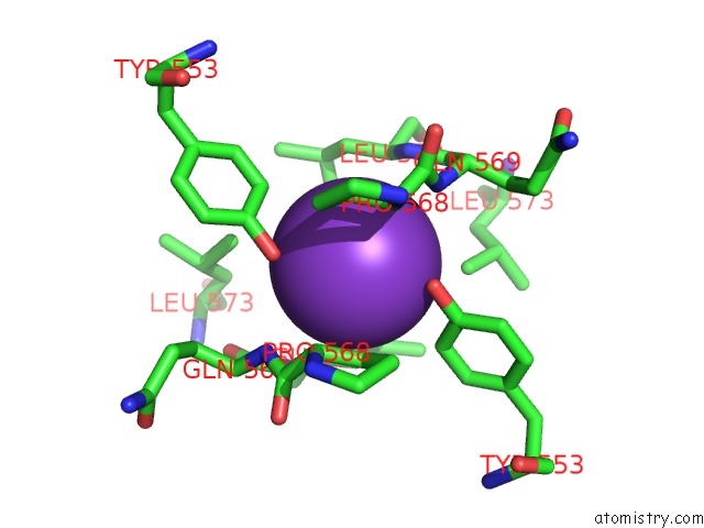
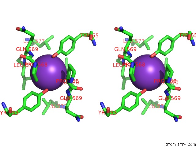
Potassium binding site 1 out of 3 in 6vc7
Go back to
Potassium binding site 1 out
of 3 in the Structure of the F349A Mutant of the Periplasmic Domain of Yejm From Salmonella Typhimurium
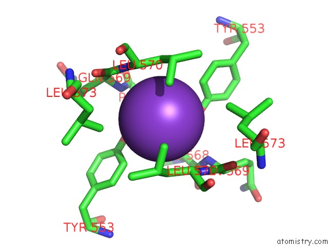
Mono view
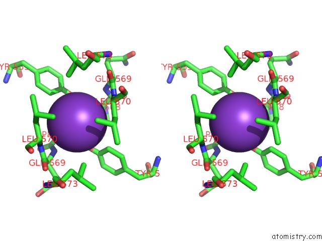
Stereo pair view
Mono view
Stereo pair view
A full contact list of Potassium with other atoms in the K binding
site number 1 of Structure of the F349A Mutant of the Periplasmic Domain of Yejm From Salmonella Typhimurium within 5.0Å range:
|
Potassium binding site 2 out of 3 in 6vc7
Go back to
Potassium binding site 2 out
of 3 in the Structure of the F349A Mutant of the Periplasmic Domain of Yejm From Salmonella Typhimurium
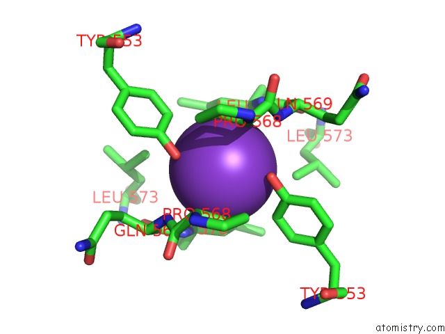
Mono view
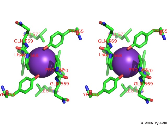
Stereo pair view
Mono view
Stereo pair view
A full contact list of Potassium with other atoms in the K binding
site number 2 of Structure of the F349A Mutant of the Periplasmic Domain of Yejm From Salmonella Typhimurium within 5.0Å range:
|
Potassium binding site 3 out of 3 in 6vc7
Go back to
Potassium binding site 3 out
of 3 in the Structure of the F349A Mutant of the Periplasmic Domain of Yejm From Salmonella Typhimurium
Mono view
Stereo pair view
Mono view
Stereo pair view
A full contact list of Potassium with other atoms in the K binding
site number 3 of Structure of the F349A Mutant of the Periplasmic Domain of Yejm From Salmonella Typhimurium within 5.0Å range:
|
Reference:
U.Gabale,
P.A.Pena Palomino,
H.Kim,
W.Chen,
S.Ressl.
The Essential Inner Membrane Protein Yejm Is A Metalloenzyme. Sci Rep V. 10 17794 2020.
ISSN: ESSN 2045-2322
PubMed: 33082366
DOI: 10.1038/S41598-020-73660-6
Page generated: Mon Aug 12 17:58:59 2024
ISSN: ESSN 2045-2322
PubMed: 33082366
DOI: 10.1038/S41598-020-73660-6
Last articles
Zn in 9J0NZn in 9J0O
Zn in 9J0P
Zn in 9FJX
Zn in 9EKB
Zn in 9C0F
Zn in 9CAH
Zn in 9CH0
Zn in 9CH3
Zn in 9CH1