Potassium »
PDB 6u9t-6v7h »
6usa »
Potassium in PDB 6usa: Crystal Structure of Tryptophan Synthase From M. Tuberculosis - Aminoacrylate- and GSK1-Bound Form
Enzymatic activity of Crystal Structure of Tryptophan Synthase From M. Tuberculosis - Aminoacrylate- and GSK1-Bound Form
All present enzymatic activity of Crystal Structure of Tryptophan Synthase From M. Tuberculosis - Aminoacrylate- and GSK1-Bound Form:
4.2.1.20;
4.2.1.20;
Protein crystallography data
The structure of Crystal Structure of Tryptophan Synthase From M. Tuberculosis - Aminoacrylate- and GSK1-Bound Form, PDB code: 6usa
was solved by
C.Chang,
K.Michalska,
N.I.Maltseva,
R.Jedrzejczak,
P.Mccarren,
P.P.Nag,
A.Joachimiak,
with X-Ray Crystallography technique. A brief refinement statistics is given in the table below:
| Resolution Low / High (Å) | 29.89 / 2.41 |
| Space group | P 21 21 21 |
| Cell size a, b, c (Å), α, β, γ (°) | 134.920, 160.037, 165.151, 90.00, 90.00, 90.00 |
| R / Rfree (%) | 15.8 / 19.2 |
Other elements in 6usa:
The structure of Crystal Structure of Tryptophan Synthase From M. Tuberculosis - Aminoacrylate- and GSK1-Bound Form also contains other interesting chemical elements:
| Chlorine | (Cl) | 4 atoms |
| Sodium | (Na) | 2 atoms |
Potassium Binding Sites:
The binding sites of Potassium atom in the Crystal Structure of Tryptophan Synthase From M. Tuberculosis - Aminoacrylate- and GSK1-Bound Form
(pdb code 6usa). This binding sites where shown within
5.0 Angstroms radius around Potassium atom.
In total 10 binding sites of Potassium where determined in the Crystal Structure of Tryptophan Synthase From M. Tuberculosis - Aminoacrylate- and GSK1-Bound Form, PDB code: 6usa:
Jump to Potassium binding site number: 1; 2; 3; 4; 5; 6; 7; 8; 9; 10;
In total 10 binding sites of Potassium where determined in the Crystal Structure of Tryptophan Synthase From M. Tuberculosis - Aminoacrylate- and GSK1-Bound Form, PDB code: 6usa:
Jump to Potassium binding site number: 1; 2; 3; 4; 5; 6; 7; 8; 9; 10;
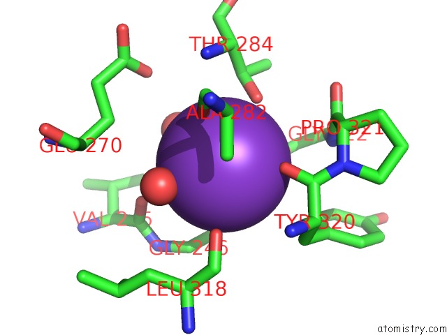
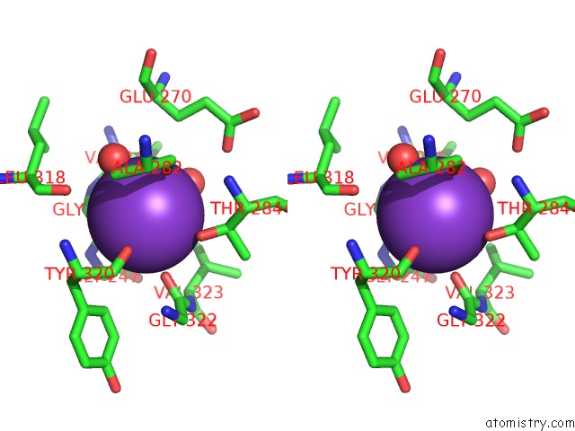
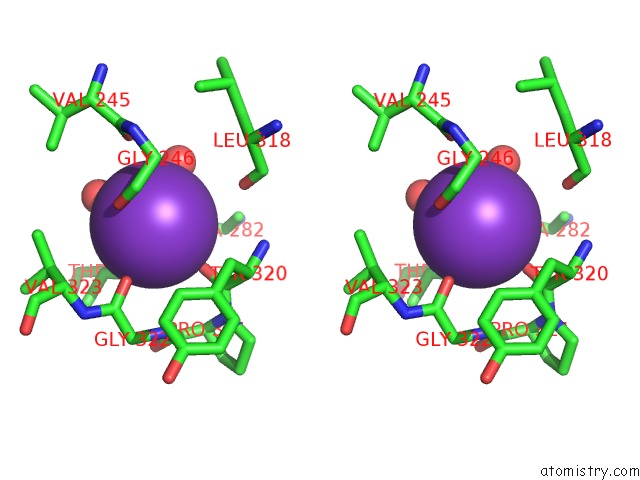
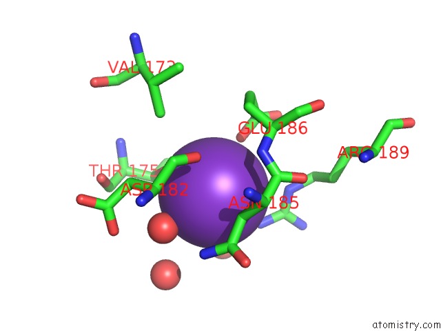
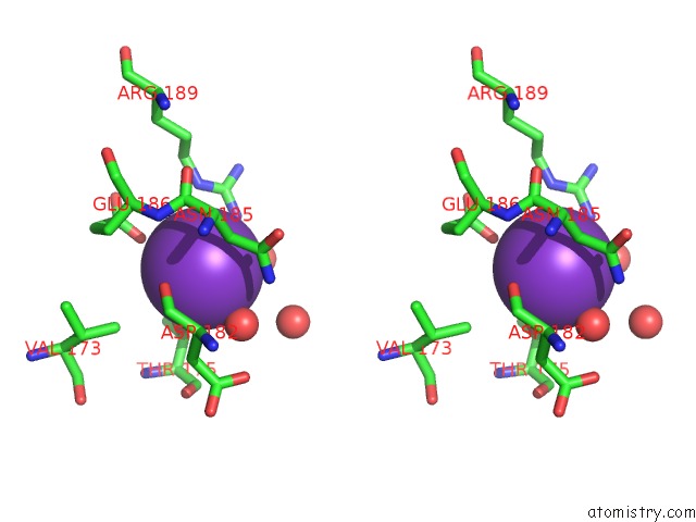
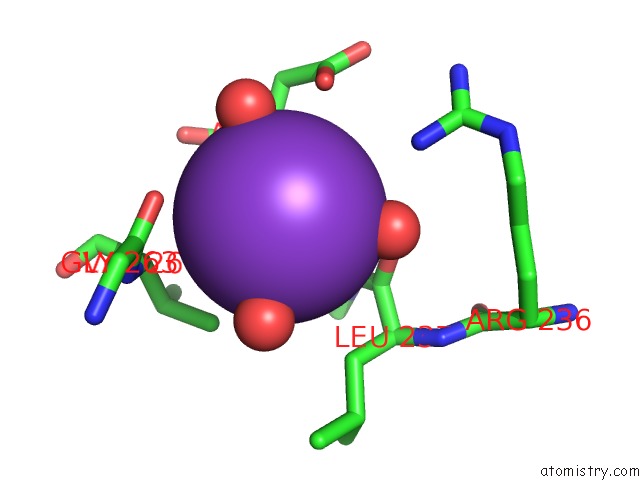
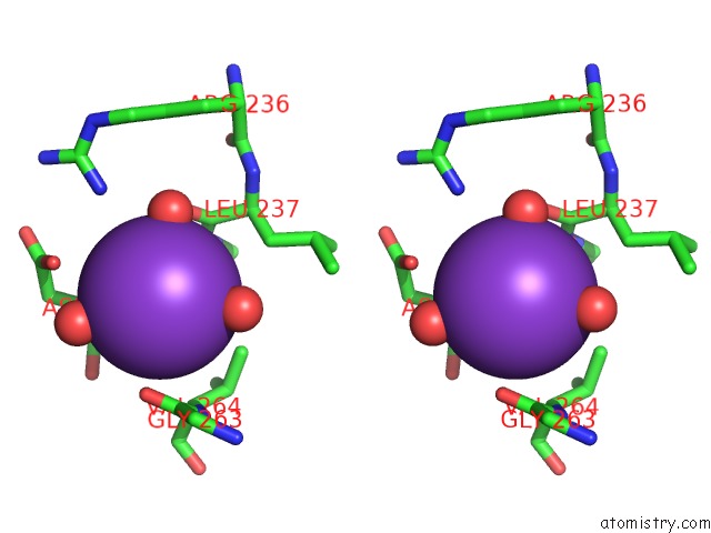
Potassium binding site 1 out of 10 in 6usa
Go back to
Potassium binding site 1 out
of 10 in the Crystal Structure of Tryptophan Synthase From M. Tuberculosis - Aminoacrylate- and GSK1-Bound Form
Mono view
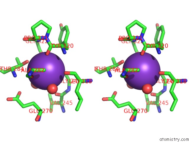
Stereo pair view
Mono view
Stereo pair view
A full contact list of Potassium with other atoms in the K binding
site number 1 of Crystal Structure of Tryptophan Synthase From M. Tuberculosis - Aminoacrylate- and GSK1-Bound Form within 5.0Å range:
|
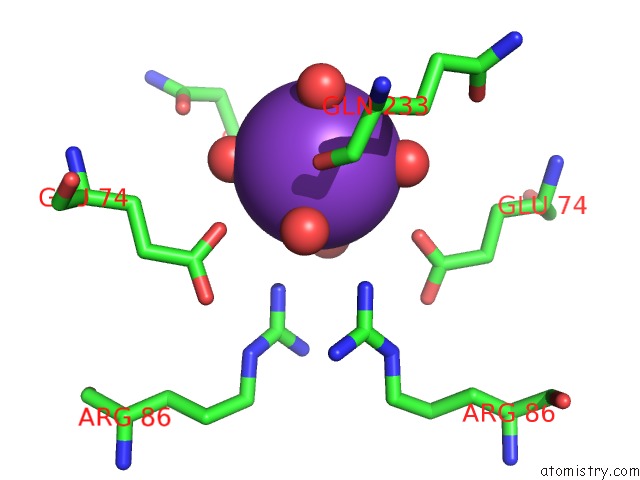
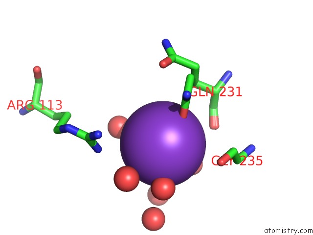
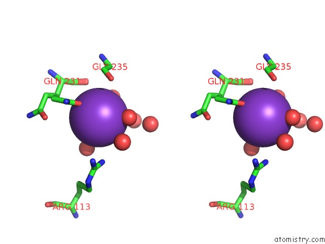
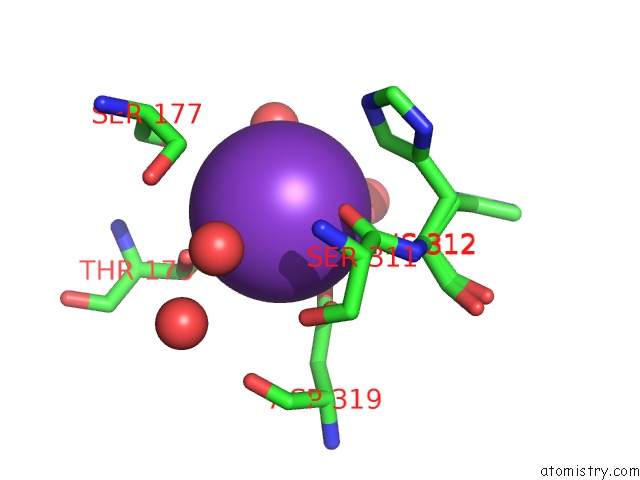
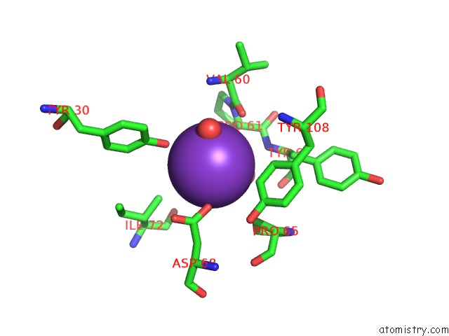
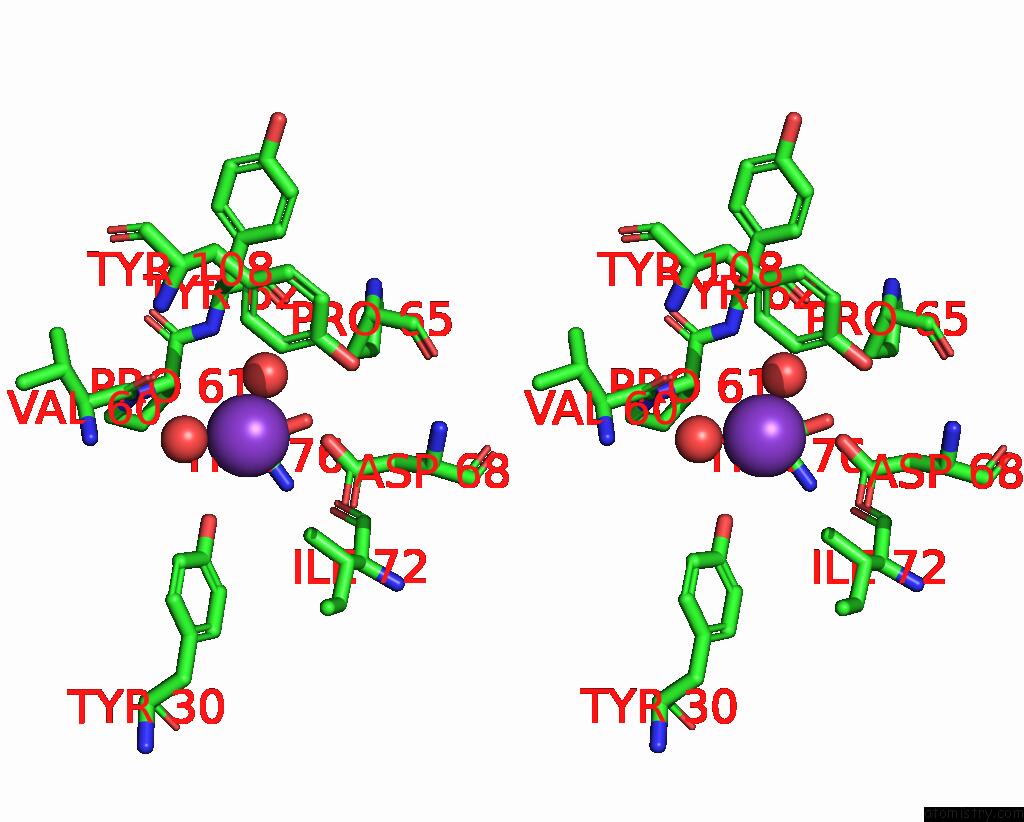
Potassium binding site 2 out of 10 in 6usa
Go back to
Potassium binding site 2 out
of 10 in the Crystal Structure of Tryptophan Synthase From M. Tuberculosis - Aminoacrylate- and GSK1-Bound Form
Mono view

Stereo pair view
Mono view
Stereo pair view
A full contact list of Potassium with other atoms in the K binding
site number 2 of Crystal Structure of Tryptophan Synthase From M. Tuberculosis - Aminoacrylate- and GSK1-Bound Form within 5.0Å range:
|
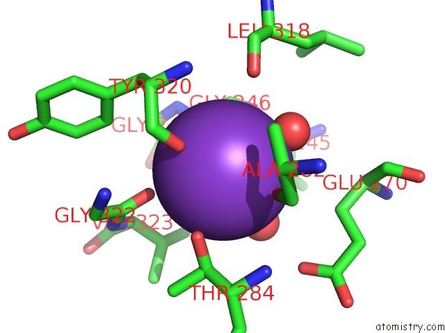
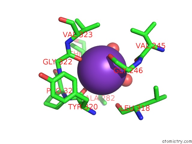
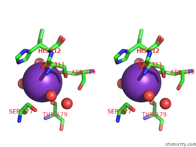
Potassium binding site 3 out of 10 in 6usa
Go back to
Potassium binding site 3 out
of 10 in the Crystal Structure of Tryptophan Synthase From M. Tuberculosis - Aminoacrylate- and GSK1-Bound Form
Mono view
Stereo pair view
Mono view
Stereo pair view
A full contact list of Potassium with other atoms in the K binding
site number 3 of Crystal Structure of Tryptophan Synthase From M. Tuberculosis - Aminoacrylate- and GSK1-Bound Form within 5.0Å range:
|
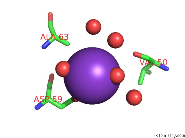
Potassium binding site 4 out of 10 in 6usa
Go back to
Potassium binding site 4 out
of 10 in the Crystal Structure of Tryptophan Synthase From M. Tuberculosis - Aminoacrylate- and GSK1-Bound Form
Mono view
Stereo pair view
Mono view
Stereo pair view
A full contact list of Potassium with other atoms in the K binding
site number 4 of Crystal Structure of Tryptophan Synthase From M. Tuberculosis - Aminoacrylate- and GSK1-Bound Form within 5.0Å range:
|
Potassium binding site 5 out of 10 in 6usa
Go back to
Potassium binding site 5 out
of 10 in the Crystal Structure of Tryptophan Synthase From M. Tuberculosis - Aminoacrylate- and GSK1-Bound Form
Mono view
Stereo pair view
Mono view
Stereo pair view
A full contact list of Potassium with other atoms in the K binding
site number 5 of Crystal Structure of Tryptophan Synthase From M. Tuberculosis - Aminoacrylate- and GSK1-Bound Form within 5.0Å range:
|
Potassium binding site 6 out of 10 in 6usa
Go back to
Potassium binding site 6 out
of 10 in the Crystal Structure of Tryptophan Synthase From M. Tuberculosis - Aminoacrylate- and GSK1-Bound Form
Mono view
Stereo pair view
Mono view
Stereo pair view
A full contact list of Potassium with other atoms in the K binding
site number 6 of Crystal Structure of Tryptophan Synthase From M. Tuberculosis - Aminoacrylate- and GSK1-Bound Form within 5.0Å range:
|
Potassium binding site 7 out of 10 in 6usa
Go back to
Potassium binding site 7 out
of 10 in the Crystal Structure of Tryptophan Synthase From M. Tuberculosis - Aminoacrylate- and GSK1-Bound Form
Mono view

Stereo pair view
Mono view
Stereo pair view
A full contact list of Potassium with other atoms in the K binding
site number 7 of Crystal Structure of Tryptophan Synthase From M. Tuberculosis - Aminoacrylate- and GSK1-Bound Form within 5.0Å range:
|
Potassium binding site 8 out of 10 in 6usa
Go back to
Potassium binding site 8 out
of 10 in the Crystal Structure of Tryptophan Synthase From M. Tuberculosis - Aminoacrylate- and GSK1-Bound Form
Mono view
Stereo pair view
Mono view
Stereo pair view
A full contact list of Potassium with other atoms in the K binding
site number 8 of Crystal Structure of Tryptophan Synthase From M. Tuberculosis - Aminoacrylate- and GSK1-Bound Form within 5.0Å range:
|
Potassium binding site 9 out of 10 in 6usa
Go back to
Potassium binding site 9 out
of 10 in the Crystal Structure of Tryptophan Synthase From M. Tuberculosis - Aminoacrylate- and GSK1-Bound Form
Mono view
Stereo pair view
Mono view
Stereo pair view
A full contact list of Potassium with other atoms in the K binding
site number 9 of Crystal Structure of Tryptophan Synthase From M. Tuberculosis - Aminoacrylate- and GSK1-Bound Form within 5.0Å range:
|
Potassium binding site 10 out of 10 in 6usa
Go back to
Potassium binding site 10 out
of 10 in the Crystal Structure of Tryptophan Synthase From M. Tuberculosis - Aminoacrylate- and GSK1-Bound Form
Mono view
Stereo pair view
Mono view
Stereo pair view
A full contact list of Potassium with other atoms in the K binding
site number 10 of Crystal Structure of Tryptophan Synthase From M. Tuberculosis - Aminoacrylate- and GSK1-Bound Form within 5.0Å range:
|
Reference:
K.Michalska,
C.Chang,
N.I.Maltseva,
R.Jedrzejczak,
G.T.Robertson,
F.Gusovsky,
P.Mccarren,
S.L.Schreiber,
P.P.Nag,
A.Joachimiak.
Allosteric Inhibitors of Mycobacterium Tuberculosis Tryptophan Synthase. Protein Sci. V. 29 779 2020.
ISSN: ESSN 1469-896X
PubMed: 31930594
DOI: 10.1002/PRO.3825
Page generated: Mon Aug 12 17:52:40 2024
ISSN: ESSN 1469-896X
PubMed: 31930594
DOI: 10.1002/PRO.3825
Last articles
Zn in 9J0NZn in 9J0O
Zn in 9J0P
Zn in 9FJX
Zn in 9EKB
Zn in 9C0F
Zn in 9CAH
Zn in 9CH0
Zn in 9CH3
Zn in 9CH1