Potassium »
PDB 6u9t-6v7h »
6uoc »
Potassium in PDB 6uoc: Crystal Structure of Danio Rerio Histone Deacetylase 6 Catalytic Domain 1 (CD1) K330L Mutant Complexed with Givinostat
Protein crystallography data
The structure of Crystal Structure of Danio Rerio Histone Deacetylase 6 Catalytic Domain 1 (CD1) K330L Mutant Complexed with Givinostat, PDB code: 6uoc
was solved by
J.D.Osko,
D.W.Christianson,
with X-Ray Crystallography technique. A brief refinement statistics is given in the table below:
| Resolution Low / High (Å) | 42.95 / 1.40 |
| Space group | P 21 21 21 |
| Cell size a, b, c (Å), α, β, γ (°) | 55.160, 60.400, 122.160, 90.00, 90.00, 90.00 |
| R / Rfree (%) | 15.2 / 16.8 |
Other elements in 6uoc:
The structure of Crystal Structure of Danio Rerio Histone Deacetylase 6 Catalytic Domain 1 (CD1) K330L Mutant Complexed with Givinostat also contains other interesting chemical elements:
| Zinc | (Zn) | 1 atom |
Potassium Binding Sites:
The binding sites of Potassium atom in the Crystal Structure of Danio Rerio Histone Deacetylase 6 Catalytic Domain 1 (CD1) K330L Mutant Complexed with Givinostat
(pdb code 6uoc). This binding sites where shown within
5.0 Angstroms radius around Potassium atom.
In total 2 binding sites of Potassium where determined in the Crystal Structure of Danio Rerio Histone Deacetylase 6 Catalytic Domain 1 (CD1) K330L Mutant Complexed with Givinostat, PDB code: 6uoc:
Jump to Potassium binding site number: 1; 2;
In total 2 binding sites of Potassium where determined in the Crystal Structure of Danio Rerio Histone Deacetylase 6 Catalytic Domain 1 (CD1) K330L Mutant Complexed with Givinostat, PDB code: 6uoc:
Jump to Potassium binding site number: 1; 2;
Potassium binding site 1 out of 2 in 6uoc
Go back to
Potassium binding site 1 out
of 2 in the Crystal Structure of Danio Rerio Histone Deacetylase 6 Catalytic Domain 1 (CD1) K330L Mutant Complexed with Givinostat
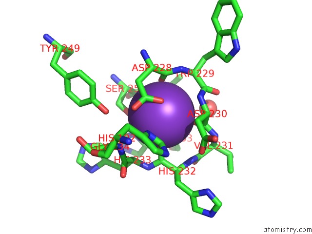
Mono view
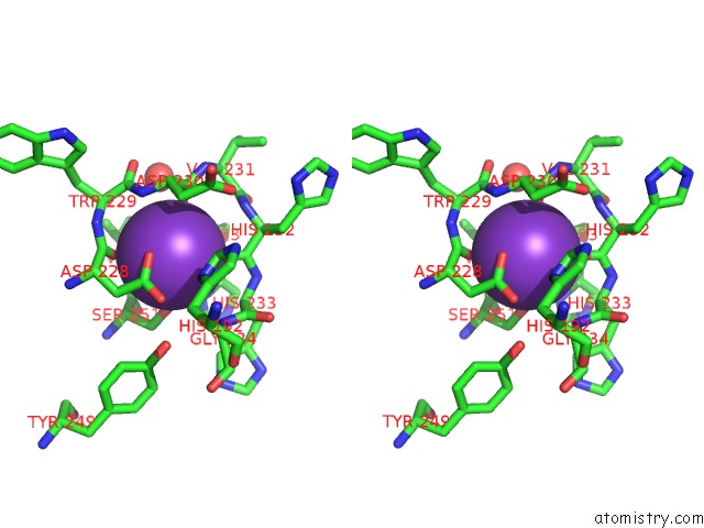
Stereo pair view
Mono view
Stereo pair view
A full contact list of Potassium with other atoms in the K binding
site number 1 of Crystal Structure of Danio Rerio Histone Deacetylase 6 Catalytic Domain 1 (CD1) K330L Mutant Complexed with Givinostat within 5.0Å range:
|
Potassium binding site 2 out of 2 in 6uoc
Go back to
Potassium binding site 2 out
of 2 in the Crystal Structure of Danio Rerio Histone Deacetylase 6 Catalytic Domain 1 (CD1) K330L Mutant Complexed with Givinostat
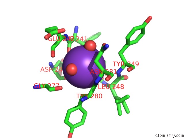
Mono view
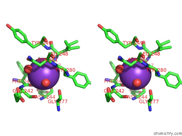
Stereo pair view
Mono view
Stereo pair view
A full contact list of Potassium with other atoms in the K binding
site number 2 of Crystal Structure of Danio Rerio Histone Deacetylase 6 Catalytic Domain 1 (CD1) K330L Mutant Complexed with Givinostat within 5.0Å range:
|
Reference:
J.D.Osko,
D.W.Christianson.
Structural Basis of Catalysis and Inhibition of HDAC6 CD1, the Enigmatic Catalytic Domain of Histone Deacetylase 6. Biochemistry 2019.
ISSN: ISSN 0006-2960
PubMed: 31755702
DOI: 10.1021/ACS.BIOCHEM.9B00934
Page generated: Mon Aug 12 17:52:09 2024
ISSN: ISSN 0006-2960
PubMed: 31755702
DOI: 10.1021/ACS.BIOCHEM.9B00934
Last articles
Zn in 9J0NZn in 9J0O
Zn in 9J0P
Zn in 9FJX
Zn in 9EKB
Zn in 9C0F
Zn in 9CAH
Zn in 9CH0
Zn in 9CH3
Zn in 9CH1