Potassium »
PDB 6rvz-6u9p »
6tmu »
Potassium in PDB 6tmu: Crystal Structure of the Chaperonin GP146 From the Bacteriophage El 2 (Pseudomonas Aeruginosa) in Presence of Atp-Befx, Crystal Form II
Protein crystallography data
The structure of Crystal Structure of the Chaperonin GP146 From the Bacteriophage El 2 (Pseudomonas Aeruginosa) in Presence of Atp-Befx, Crystal Form II, PDB code: 6tmu
was solved by
A.Bracher,
S.S.Paul,
H.Wang,
N.Wischnewski,
F.U.Hartl,
M.Hayer-Hartl,
with X-Ray Crystallography technique. A brief refinement statistics is given in the table below:
| Resolution Low / High (Å) | 30.00 / 3.54 |
| Space group | P 21 21 21 |
| Cell size a, b, c (Å), α, β, γ (°) | 145.516, 151.461, 261.347, 90.00, 90.00, 90.00 |
| R / Rfree (%) | 25 / 27.5 |
Other elements in 6tmu:
The structure of Crystal Structure of the Chaperonin GP146 From the Bacteriophage El 2 (Pseudomonas Aeruginosa) in Presence of Atp-Befx, Crystal Form II also contains other interesting chemical elements:
| Magnesium | (Mg) | 4 atoms |
Potassium Binding Sites:
The binding sites of Potassium atom in the Crystal Structure of the Chaperonin GP146 From the Bacteriophage El 2 (Pseudomonas Aeruginosa) in Presence of Atp-Befx, Crystal Form II
(pdb code 6tmu). This binding sites where shown within
5.0 Angstroms radius around Potassium atom.
In total 4 binding sites of Potassium where determined in the Crystal Structure of the Chaperonin GP146 From the Bacteriophage El 2 (Pseudomonas Aeruginosa) in Presence of Atp-Befx, Crystal Form II, PDB code: 6tmu:
Jump to Potassium binding site number: 1; 2; 3; 4;
In total 4 binding sites of Potassium where determined in the Crystal Structure of the Chaperonin GP146 From the Bacteriophage El 2 (Pseudomonas Aeruginosa) in Presence of Atp-Befx, Crystal Form II, PDB code: 6tmu:
Jump to Potassium binding site number: 1; 2; 3; 4;
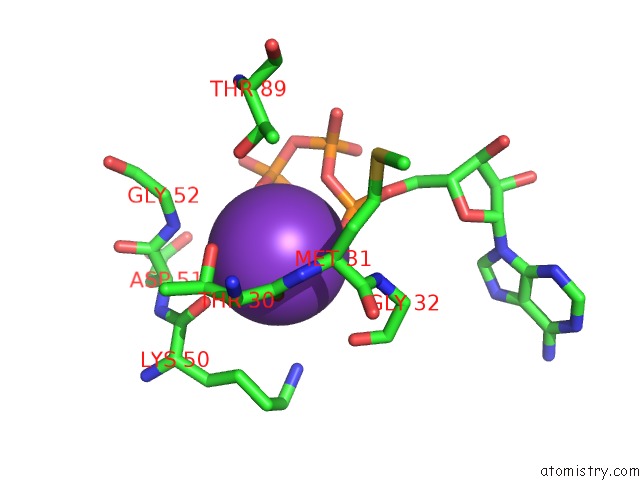
Potassium binding site 1 out of 4 in 6tmu
Go back to
Potassium binding site 1 out
of 4 in the Crystal Structure of the Chaperonin GP146 From the Bacteriophage El 2 (Pseudomonas Aeruginosa) in Presence of Atp-Befx, Crystal Form II
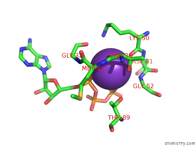
Mono view
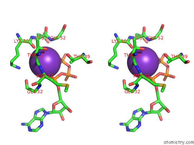
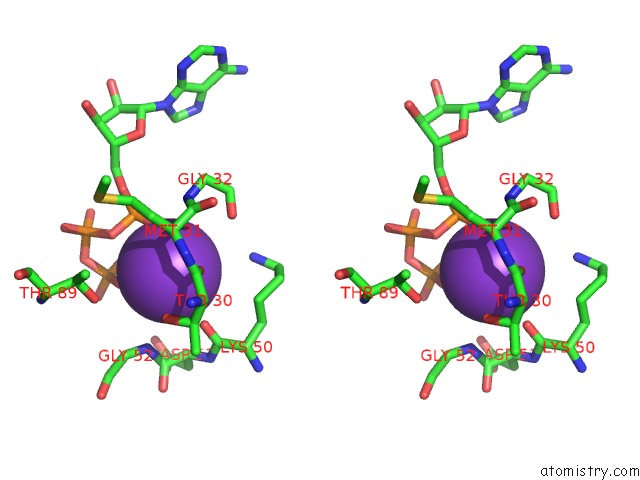
Stereo pair view
Mono view
Stereo pair view
A full contact list of Potassium with other atoms in the K binding
site number 1 of Crystal Structure of the Chaperonin GP146 From the Bacteriophage El 2 (Pseudomonas Aeruginosa) in Presence of Atp-Befx, Crystal Form II within 5.0Å range:
|
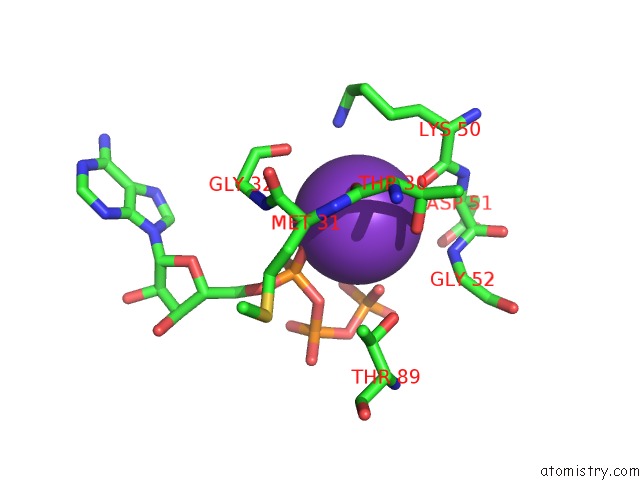
Potassium binding site 2 out of 4 in 6tmu
Go back to
Potassium binding site 2 out
of 4 in the Crystal Structure of the Chaperonin GP146 From the Bacteriophage El 2 (Pseudomonas Aeruginosa) in Presence of Atp-Befx, Crystal Form II
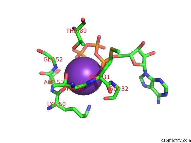
Mono view
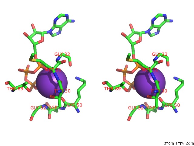
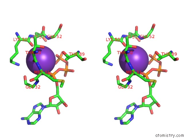
Stereo pair view
Mono view
Stereo pair view
A full contact list of Potassium with other atoms in the K binding
site number 2 of Crystal Structure of the Chaperonin GP146 From the Bacteriophage El 2 (Pseudomonas Aeruginosa) in Presence of Atp-Befx, Crystal Form II within 5.0Å range:
|
Potassium binding site 3 out of 4 in 6tmu
Go back to
Potassium binding site 3 out
of 4 in the Crystal Structure of the Chaperonin GP146 From the Bacteriophage El 2 (Pseudomonas Aeruginosa) in Presence of Atp-Befx, Crystal Form II
Mono view
Stereo pair view
Mono view
Stereo pair view
A full contact list of Potassium with other atoms in the K binding
site number 3 of Crystal Structure of the Chaperonin GP146 From the Bacteriophage El 2 (Pseudomonas Aeruginosa) in Presence of Atp-Befx, Crystal Form II within 5.0Å range:
|
Potassium binding site 4 out of 4 in 6tmu
Go back to
Potassium binding site 4 out
of 4 in the Crystal Structure of the Chaperonin GP146 From the Bacteriophage El 2 (Pseudomonas Aeruginosa) in Presence of Atp-Befx, Crystal Form II
Mono view
Stereo pair view
Mono view
Stereo pair view
A full contact list of Potassium with other atoms in the K binding
site number 4 of Crystal Structure of the Chaperonin GP146 From the Bacteriophage El 2 (Pseudomonas Aeruginosa) in Presence of Atp-Befx, Crystal Form II within 5.0Å range:
|
Reference:
A.Bracher,
S.S.Paul,
H.Wang,
N.Wischnewski,
F.U.Hartl,
M.Hayer-Hartl.
Structure and Conformational Cycle of A Bacteriophage-Encoded Chaperonin Plos One 2020.
ISSN: ESSN 1932-6203
Page generated: Sat Aug 9 12:12:36 2025
ISSN: ESSN 1932-6203
Last articles
Mg in 1INIMg in 1IBK
Mg in 1IKK
Mg in 1IK5
Mg in 1IJJ
Mg in 1IJF
Mg in 1IJD
Mg in 1IIR
Mg in 1II9
Mg in 1II6