Potassium »
PDB 6rvz-6u9p »
6sbi »
Potassium in PDB 6sbi: X-Ray Structure of Murine Fumarylacetoacetate Hydrolase Domain Containing Protein 1 (FAHD1) in Complex with Inhibitor Oxalate
Enzymatic activity of X-Ray Structure of Murine Fumarylacetoacetate Hydrolase Domain Containing Protein 1 (FAHD1) in Complex with Inhibitor Oxalate
All present enzymatic activity of X-Ray Structure of Murine Fumarylacetoacetate Hydrolase Domain Containing Protein 1 (FAHD1) in Complex with Inhibitor Oxalate:
3.7.1.5; 4.1.1.112;
3.7.1.5; 4.1.1.112;
Protein crystallography data
The structure of X-Ray Structure of Murine Fumarylacetoacetate Hydrolase Domain Containing Protein 1 (FAHD1) in Complex with Inhibitor Oxalate, PDB code: 6sbi
was solved by
B.Rupp,
A.Naschberger,
A.K.H.Weiss,
with X-Ray Crystallography technique. A brief refinement statistics is given in the table below:
| Resolution Low / High (Å) | 44.37 / 2.70 |
| Space group | P 1 21 1 |
| Cell size a, b, c (Å), α, β, γ (°) | 52.603, 103.348, 86.597, 90.00, 90.21, 90.00 |
| R / Rfree (%) | 18.7 / 23.4 |
Other elements in 6sbi:
The structure of X-Ray Structure of Murine Fumarylacetoacetate Hydrolase Domain Containing Protein 1 (FAHD1) in Complex with Inhibitor Oxalate also contains other interesting chemical elements:
| Magnesium | (Mg) | 4 atoms |
| Chlorine | (Cl) | 9 atoms |
Potassium Binding Sites:
The binding sites of Potassium atom in the X-Ray Structure of Murine Fumarylacetoacetate Hydrolase Domain Containing Protein 1 (FAHD1) in Complex with Inhibitor Oxalate
(pdb code 6sbi). This binding sites where shown within
5.0 Angstroms radius around Potassium atom.
In total 2 binding sites of Potassium where determined in the X-Ray Structure of Murine Fumarylacetoacetate Hydrolase Domain Containing Protein 1 (FAHD1) in Complex with Inhibitor Oxalate, PDB code: 6sbi:
Jump to Potassium binding site number: 1; 2;
In total 2 binding sites of Potassium where determined in the X-Ray Structure of Murine Fumarylacetoacetate Hydrolase Domain Containing Protein 1 (FAHD1) in Complex with Inhibitor Oxalate, PDB code: 6sbi:
Jump to Potassium binding site number: 1; 2;
Potassium binding site 1 out of 2 in 6sbi
Go back to
Potassium binding site 1 out
of 2 in the X-Ray Structure of Murine Fumarylacetoacetate Hydrolase Domain Containing Protein 1 (FAHD1) in Complex with Inhibitor Oxalate
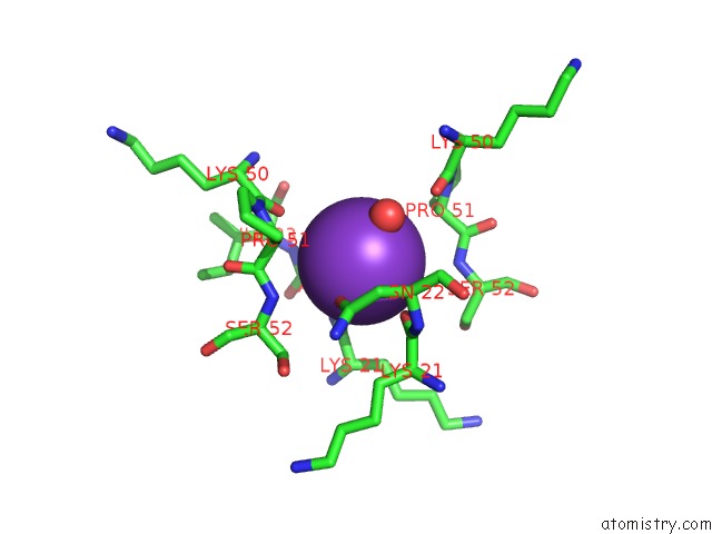
Mono view
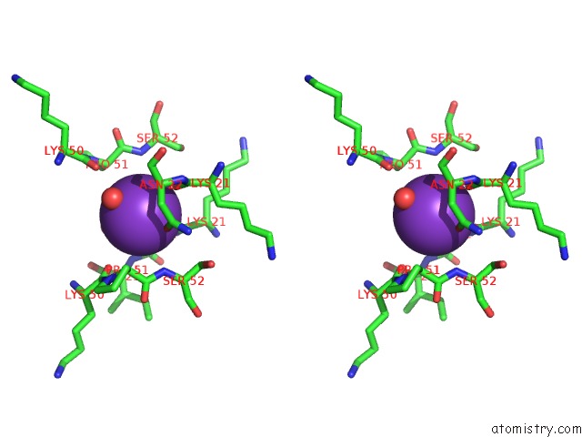
Stereo pair view
Mono view
Stereo pair view
A full contact list of Potassium with other atoms in the K binding
site number 1 of X-Ray Structure of Murine Fumarylacetoacetate Hydrolase Domain Containing Protein 1 (FAHD1) in Complex with Inhibitor Oxalate within 5.0Å range:
|
Potassium binding site 2 out of 2 in 6sbi
Go back to
Potassium binding site 2 out
of 2 in the X-Ray Structure of Murine Fumarylacetoacetate Hydrolase Domain Containing Protein 1 (FAHD1) in Complex with Inhibitor Oxalate
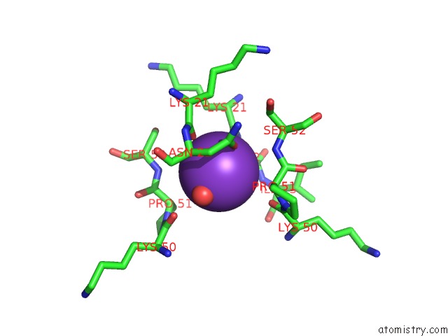
Mono view
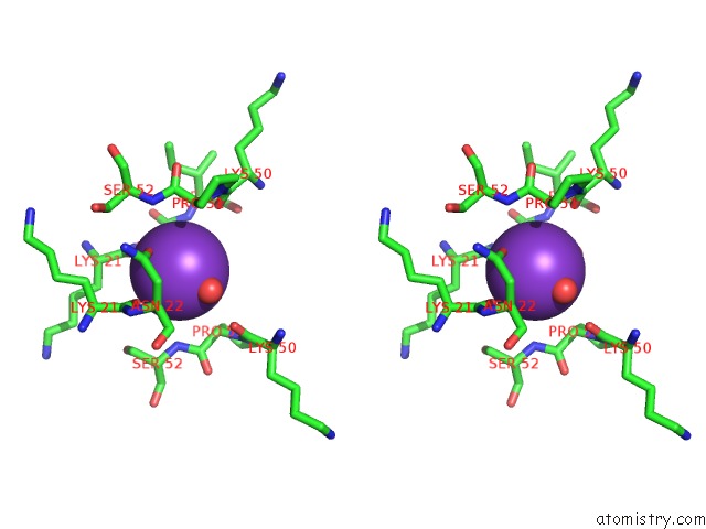
Stereo pair view
Mono view
Stereo pair view
A full contact list of Potassium with other atoms in the K binding
site number 2 of X-Ray Structure of Murine Fumarylacetoacetate Hydrolase Domain Containing Protein 1 (FAHD1) in Complex with Inhibitor Oxalate within 5.0Å range:
|
Reference:
A.K.H.Weiss,
A.Naschberger,
E.Cappuccio,
C.Metzger,
L.Mottes,
M.Holzknecht,
J.Von Velsen,
M.W.Bowler,
B.Rupp,
P.Jansen-Durr.
Structural and Functional Comparison of Fumarylacetoacetate Domain Containing Protein 1 in Human and Mouse. Biosci.Rep. V. 40 2020.
ISSN: ISSN 0144-8463
PubMed: 32068790
DOI: 10.1042/BSR20194431
Page generated: Mon Aug 12 17:35:03 2024
ISSN: ISSN 0144-8463
PubMed: 32068790
DOI: 10.1042/BSR20194431
Last articles
Zn in 9MJ5Zn in 9HNW
Zn in 9G0L
Zn in 9FNE
Zn in 9DZN
Zn in 9E0I
Zn in 9D32
Zn in 9DAK
Zn in 8ZXC
Zn in 8ZUF