Potassium »
PDB 6rvz-6u9p »
6rwv »
Potassium in PDB 6rwv: Structure of Apo-Lmcpfc
Enzymatic activity of Structure of Apo-Lmcpfc
All present enzymatic activity of Structure of Apo-Lmcpfc:
4.99.1.1;
4.99.1.1;
Protein crystallography data
The structure of Structure of Apo-Lmcpfc, PDB code: 6rwv
was solved by
S.Hofbauer,
J.Helm,
K.Djinovic-Carugo,
P.G.Furtmueller,
with X-Ray Crystallography technique. A brief refinement statistics is given in the table below:
| Resolution Low / High (Å) | 50.09 / 1.64 |
| Space group | P 1 21 1 |
| Cell size a, b, c (Å), α, β, γ (°) | 48.303, 76.723, 52.258, 90.00, 106.56, 90.00 |
| R / Rfree (%) | 17.6 / 20.1 |
Potassium Binding Sites:
The binding sites of Potassium atom in the Structure of Apo-Lmcpfc
(pdb code 6rwv). This binding sites where shown within
5.0 Angstroms radius around Potassium atom.
In total 2 binding sites of Potassium where determined in the Structure of Apo-Lmcpfc, PDB code: 6rwv:
Jump to Potassium binding site number: 1; 2;
In total 2 binding sites of Potassium where determined in the Structure of Apo-Lmcpfc, PDB code: 6rwv:
Jump to Potassium binding site number: 1; 2;
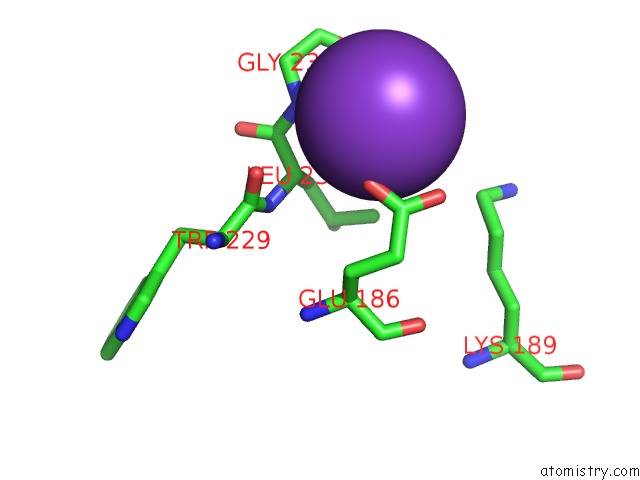
Potassium binding site 1 out of 2 in 6rwv
Go back to
Potassium binding site 1 out
of 2 in the Structure of Apo-Lmcpfc
Mono view

Stereo pair view
Mono view
Stereo pair view
A full contact list of Potassium with other atoms in the K binding
site number 1 of Structure of Apo-Lmcpfc within 5.0Å range:
|
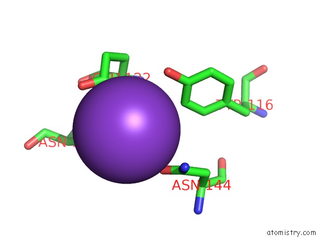
Potassium binding site 2 out of 2 in 6rwv
Go back to
Potassium binding site 2 out
of 2 in the Structure of Apo-Lmcpfc
Mono view

Stereo pair view
Mono view
Stereo pair view
A full contact list of Potassium with other atoms in the K binding
site number 2 of Structure of Apo-Lmcpfc within 5.0Å range:
|
Reference:
S.Hofbauer,
J.Helm,
C.Obinger,
K.Djinovic-Carugo,
P.G.Furtmuller.
Crystal Structures and Calorimetry Reveal Catalytically Relevant Binding Mode of Coproporphyrin and Coproheme in Coproporphyrin Ferrochelatase. Febs J. 2019.
ISSN: ISSN 1742-464X
PubMed: 31794133
DOI: 10.1111/FEBS.15164
Page generated: Mon Aug 12 17:34:14 2024
ISSN: ISSN 1742-464X
PubMed: 31794133
DOI: 10.1111/FEBS.15164
Last articles
Zn in 9J0NZn in 9J0O
Zn in 9J0P
Zn in 9FJX
Zn in 9EKB
Zn in 9C0F
Zn in 9CAH
Zn in 9CH0
Zn in 9CH3
Zn in 9CH1