Potassium »
PDB 6pcd-6qm2 »
6q6r »
Potassium in PDB 6q6r: Recognition of Different Base Tetrads By Rhau: X-Ray Crystal Structure of G4 Recognition Motif Bound to the 3-End Tetrad of A Dna G- Quadruplex
Enzymatic activity of Recognition of Different Base Tetrads By Rhau: X-Ray Crystal Structure of G4 Recognition Motif Bound to the 3-End Tetrad of A Dna G- Quadruplex
All present enzymatic activity of Recognition of Different Base Tetrads By Rhau: X-Ray Crystal Structure of G4 Recognition Motif Bound to the 3-End Tetrad of A Dna G- Quadruplex:
3.6.4.12; 3.6.4.13;
3.6.4.12; 3.6.4.13;
Protein crystallography data
The structure of Recognition of Different Base Tetrads By Rhau: X-Ray Crystal Structure of G4 Recognition Motif Bound to the 3-End Tetrad of A Dna G- Quadruplex, PDB code: 6q6r
was solved by
B.Heddi,
V.V.Cheong,
E.Schmitt,
Y.Mechulam,
A.T.Phan,
with X-Ray Crystallography technique. A brief refinement statistics is given in the table below:
| Resolution Low / High (Å) | 60.60 / 1.50 |
| Space group | P 1 21 1 |
| Cell size a, b, c (Å), α, β, γ (°) | 56.390, 42.250, 61.404, 90.00, 99.30, 90.00 |
| R / Rfree (%) | 18.5 / 24.5 |
Potassium Binding Sites:
The binding sites of Potassium atom in the Recognition of Different Base Tetrads By Rhau: X-Ray Crystal Structure of G4 Recognition Motif Bound to the 3-End Tetrad of A Dna G- Quadruplex
(pdb code 6q6r). This binding sites where shown within
5.0 Angstroms radius around Potassium atom.
In total 10 binding sites of Potassium where determined in the Recognition of Different Base Tetrads By Rhau: X-Ray Crystal Structure of G4 Recognition Motif Bound to the 3-End Tetrad of A Dna G- Quadruplex, PDB code: 6q6r:
Jump to Potassium binding site number: 1; 2; 3; 4; 5; 6; 7; 8; 9; 10;
In total 10 binding sites of Potassium where determined in the Recognition of Different Base Tetrads By Rhau: X-Ray Crystal Structure of G4 Recognition Motif Bound to the 3-End Tetrad of A Dna G- Quadruplex, PDB code: 6q6r:
Jump to Potassium binding site number: 1; 2; 3; 4; 5; 6; 7; 8; 9; 10;
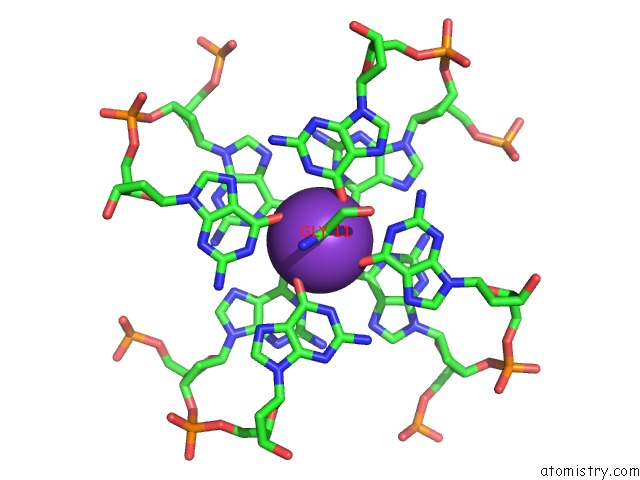
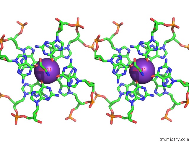
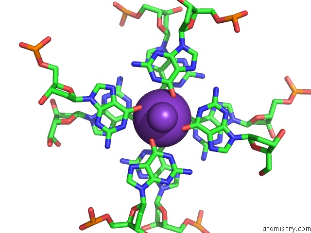
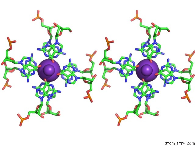
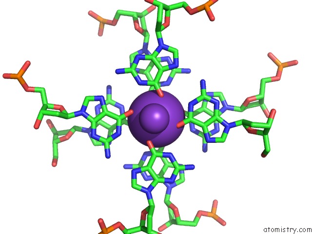
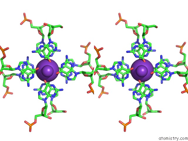
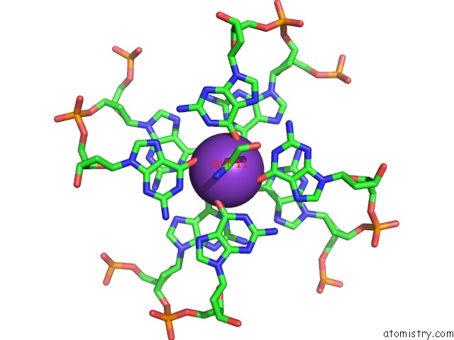
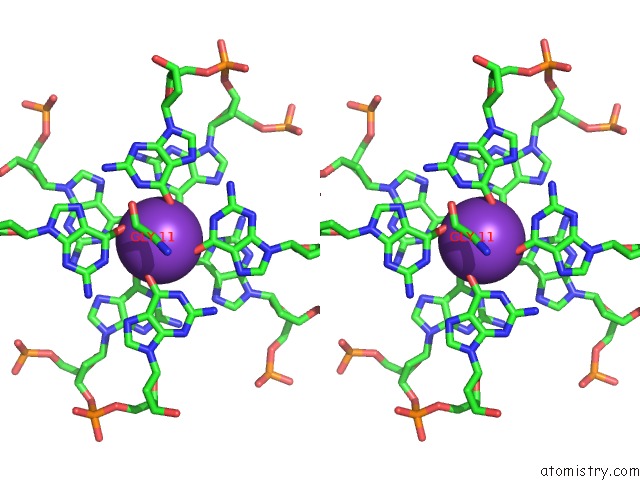
Potassium binding site 1 out of 10 in 6q6r
Go back to
Potassium binding site 1 out
of 10 in the Recognition of Different Base Tetrads By Rhau: X-Ray Crystal Structure of G4 Recognition Motif Bound to the 3-End Tetrad of A Dna G- Quadruplex
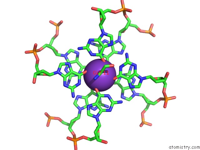
Mono view
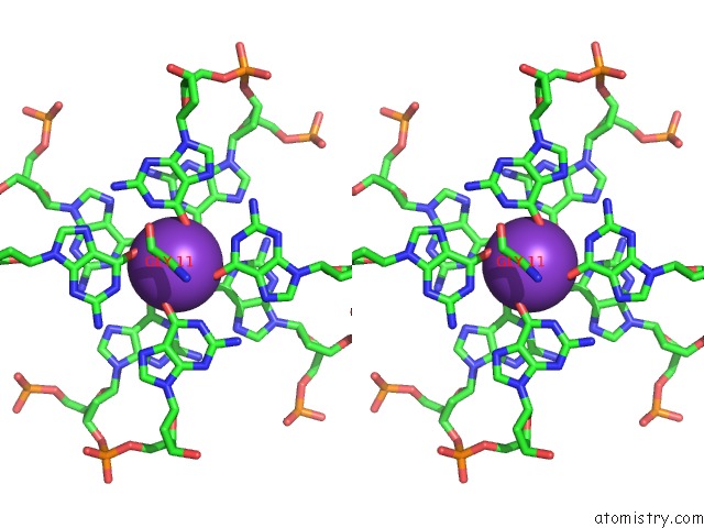
Stereo pair view
Mono view
Stereo pair view
A full contact list of Potassium with other atoms in the K binding
site number 1 of Recognition of Different Base Tetrads By Rhau: X-Ray Crystal Structure of G4 Recognition Motif Bound to the 3-End Tetrad of A Dna G- Quadruplex within 5.0Å range:
|
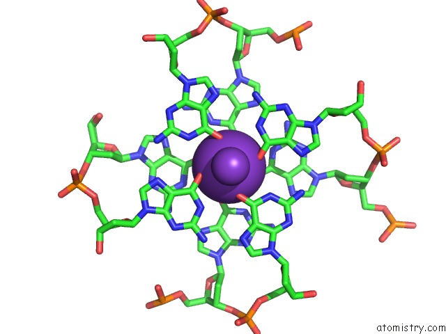
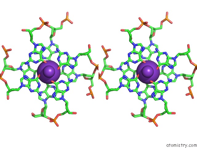
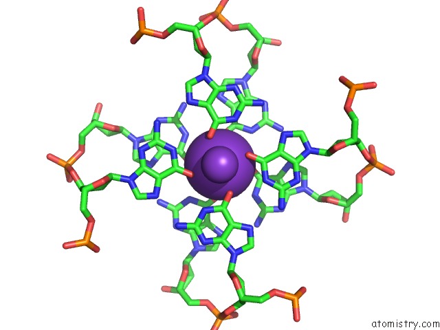
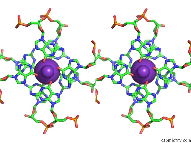
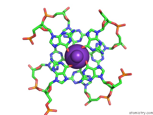
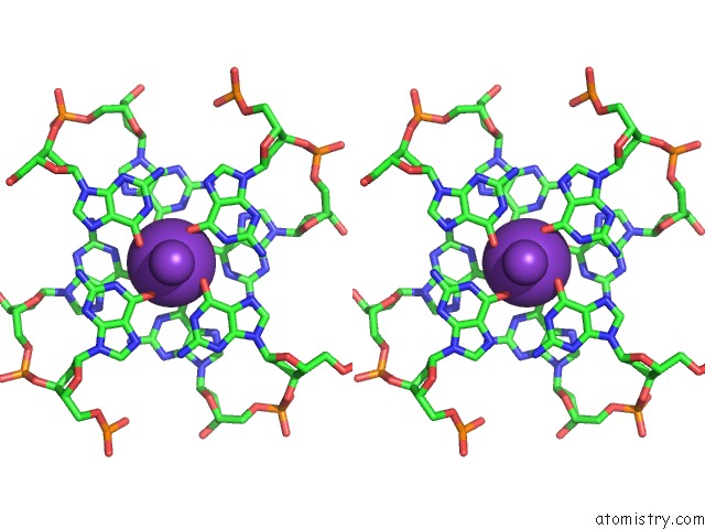
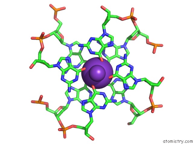
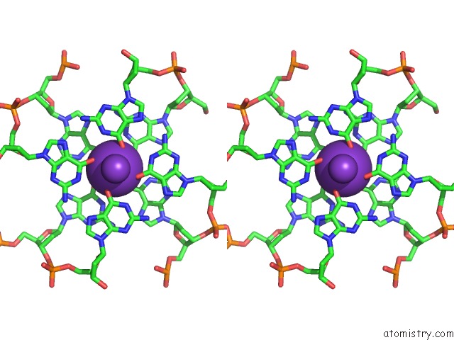
Potassium binding site 2 out of 10 in 6q6r
Go back to
Potassium binding site 2 out
of 10 in the Recognition of Different Base Tetrads By Rhau: X-Ray Crystal Structure of G4 Recognition Motif Bound to the 3-End Tetrad of A Dna G- Quadruplex
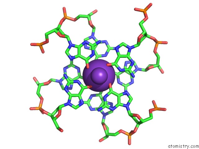
Mono view
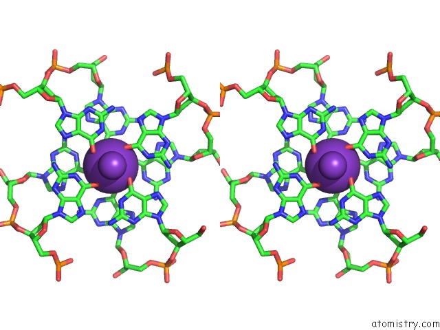
Stereo pair view
Mono view
Stereo pair view
A full contact list of Potassium with other atoms in the K binding
site number 2 of Recognition of Different Base Tetrads By Rhau: X-Ray Crystal Structure of G4 Recognition Motif Bound to the 3-End Tetrad of A Dna G- Quadruplex within 5.0Å range:
|
Potassium binding site 3 out of 10 in 6q6r
Go back to
Potassium binding site 3 out
of 10 in the Recognition of Different Base Tetrads By Rhau: X-Ray Crystal Structure of G4 Recognition Motif Bound to the 3-End Tetrad of A Dna G- Quadruplex
Mono view
Stereo pair view
Mono view
Stereo pair view
A full contact list of Potassium with other atoms in the K binding
site number 3 of Recognition of Different Base Tetrads By Rhau: X-Ray Crystal Structure of G4 Recognition Motif Bound to the 3-End Tetrad of A Dna G- Quadruplex within 5.0Å range:
|
Potassium binding site 4 out of 10 in 6q6r
Go back to
Potassium binding site 4 out
of 10 in the Recognition of Different Base Tetrads By Rhau: X-Ray Crystal Structure of G4 Recognition Motif Bound to the 3-End Tetrad of A Dna G- Quadruplex
Mono view
Stereo pair view
Mono view
Stereo pair view
A full contact list of Potassium with other atoms in the K binding
site number 4 of Recognition of Different Base Tetrads By Rhau: X-Ray Crystal Structure of G4 Recognition Motif Bound to the 3-End Tetrad of A Dna G- Quadruplex within 5.0Å range:
|
Potassium binding site 5 out of 10 in 6q6r
Go back to
Potassium binding site 5 out
of 10 in the Recognition of Different Base Tetrads By Rhau: X-Ray Crystal Structure of G4 Recognition Motif Bound to the 3-End Tetrad of A Dna G- Quadruplex
Mono view
Stereo pair view
Mono view
Stereo pair view
A full contact list of Potassium with other atoms in the K binding
site number 5 of Recognition of Different Base Tetrads By Rhau: X-Ray Crystal Structure of G4 Recognition Motif Bound to the 3-End Tetrad of A Dna G- Quadruplex within 5.0Å range:
|
Potassium binding site 6 out of 10 in 6q6r
Go back to
Potassium binding site 6 out
of 10 in the Recognition of Different Base Tetrads By Rhau: X-Ray Crystal Structure of G4 Recognition Motif Bound to the 3-End Tetrad of A Dna G- Quadruplex
Mono view
Stereo pair view
Mono view
Stereo pair view
A full contact list of Potassium with other atoms in the K binding
site number 6 of Recognition of Different Base Tetrads By Rhau: X-Ray Crystal Structure of G4 Recognition Motif Bound to the 3-End Tetrad of A Dna G- Quadruplex within 5.0Å range:
|
Potassium binding site 7 out of 10 in 6q6r
Go back to
Potassium binding site 7 out
of 10 in the Recognition of Different Base Tetrads By Rhau: X-Ray Crystal Structure of G4 Recognition Motif Bound to the 3-End Tetrad of A Dna G- Quadruplex
Mono view
Stereo pair view
Mono view
Stereo pair view
A full contact list of Potassium with other atoms in the K binding
site number 7 of Recognition of Different Base Tetrads By Rhau: X-Ray Crystal Structure of G4 Recognition Motif Bound to the 3-End Tetrad of A Dna G- Quadruplex within 5.0Å range:
|
Potassium binding site 8 out of 10 in 6q6r
Go back to
Potassium binding site 8 out
of 10 in the Recognition of Different Base Tetrads By Rhau: X-Ray Crystal Structure of G4 Recognition Motif Bound to the 3-End Tetrad of A Dna G- Quadruplex
Mono view
Stereo pair view
Mono view
Stereo pair view
A full contact list of Potassium with other atoms in the K binding
site number 8 of Recognition of Different Base Tetrads By Rhau: X-Ray Crystal Structure of G4 Recognition Motif Bound to the 3-End Tetrad of A Dna G- Quadruplex within 5.0Å range:
|
Potassium binding site 9 out of 10 in 6q6r
Go back to
Potassium binding site 9 out
of 10 in the Recognition of Different Base Tetrads By Rhau: X-Ray Crystal Structure of G4 Recognition Motif Bound to the 3-End Tetrad of A Dna G- Quadruplex
Mono view
Stereo pair view
Mono view
Stereo pair view
A full contact list of Potassium with other atoms in the K binding
site number 9 of Recognition of Different Base Tetrads By Rhau: X-Ray Crystal Structure of G4 Recognition Motif Bound to the 3-End Tetrad of A Dna G- Quadruplex within 5.0Å range:
|
Potassium binding site 10 out of 10 in 6q6r
Go back to
Potassium binding site 10 out
of 10 in the Recognition of Different Base Tetrads By Rhau: X-Ray Crystal Structure of G4 Recognition Motif Bound to the 3-End Tetrad of A Dna G- Quadruplex
Mono view
Stereo pair view
Mono view
Stereo pair view
A full contact list of Potassium with other atoms in the K binding
site number 10 of Recognition of Different Base Tetrads By Rhau: X-Ray Crystal Structure of G4 Recognition Motif Bound to the 3-End Tetrad of A Dna G- Quadruplex within 5.0Å range:
|
Reference:
B.Heddi,
V.V.Cheong,
E.Schmitt,
Y.Mechulam,
A.T.Phan.
Recognition of Different Base Tetrads By Rhau (DHX36): X-Ray Crystal Structure of the G4 Recognition Motif Bound to the 3'-End Tetrad of A Dna G-Quadruplex. J.Struct.Biol. 07399 2019.
ISSN: ESSN 1095-8657
PubMed: 31586599
DOI: 10.1016/J.JSB.2019.10.001
Page generated: Mon Aug 12 17:17:12 2024
ISSN: ESSN 1095-8657
PubMed: 31586599
DOI: 10.1016/J.JSB.2019.10.001
Last articles
Zn in 9J0NZn in 9J0O
Zn in 9J0P
Zn in 9FJX
Zn in 9EKB
Zn in 9C0F
Zn in 9CAH
Zn in 9CH0
Zn in 9CH3
Zn in 9CH1