Potassium »
PDB 6pcd-6qm2 »
6pnk »
Potassium in PDB 6pnk: Crystal Structure of the G-Quadruplex Formed By (Gggtt)3GGG in Complex with N-Methylmesoporphryin IX
Protein crystallography data
The structure of Crystal Structure of the G-Quadruplex Formed By (Gggtt)3GGG in Complex with N-Methylmesoporphryin IX, PDB code: 6pnk
was solved by
L.A.Yatsunyk,
L.Y.Lin,
with X-Ray Crystallography technique. A brief refinement statistics is given in the table below:
| Resolution Low / High (Å) | 64.78 / 2.39 |
| Space group | H 3 2 |
| Cell size a, b, c (Å), α, β, γ (°) | 60.930, 60.930, 194.342, 90.00, 90.00, 120.00 |
| R / Rfree (%) | 23.5 / 24.9 |
Other elements in 6pnk:
The structure of Crystal Structure of the G-Quadruplex Formed By (Gggtt)3GGG in Complex with N-Methylmesoporphryin IX also contains other interesting chemical elements:
| Sodium | (Na) | 1 atom |
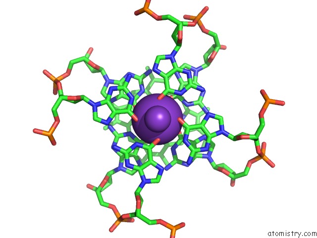
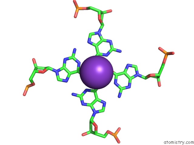
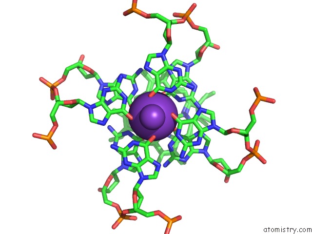
Potassium Binding Sites:
The binding sites of Potassium atom in the Crystal Structure of the G-Quadruplex Formed By (Gggtt)3GGG in Complex with N-Methylmesoporphryin IX
(pdb code 6pnk). This binding sites where shown within
5.0 Angstroms radius around Potassium atom.
In total 6 binding sites of Potassium where determined in the Crystal Structure of the G-Quadruplex Formed By (Gggtt)3GGG in Complex with N-Methylmesoporphryin IX, PDB code: 6pnk:
Jump to Potassium binding site number: 1; 2; 3; 4; 5; 6;
In total 6 binding sites of Potassium where determined in the Crystal Structure of the G-Quadruplex Formed By (Gggtt)3GGG in Complex with N-Methylmesoporphryin IX, PDB code: 6pnk:
Jump to Potassium binding site number: 1; 2; 3; 4; 5; 6;
Potassium binding site 1 out of 6 in 6pnk
Go back to
Potassium binding site 1 out
of 6 in the Crystal Structure of the G-Quadruplex Formed By (Gggtt)3GGG in Complex with N-Methylmesoporphryin IX

Mono view

Stereo pair view
Mono view
Stereo pair view
A full contact list of Potassium with other atoms in the K binding
site number 1 of Crystal Structure of the G-Quadruplex Formed By (Gggtt)3GGG in Complex with N-Methylmesoporphryin IX within 5.0Å range:
|
Potassium binding site 2 out of 6 in 6pnk
Go back to
Potassium binding site 2 out
of 6 in the Crystal Structure of the G-Quadruplex Formed By (Gggtt)3GGG in Complex with N-Methylmesoporphryin IX
Mono view

Stereo pair view
Mono view
Stereo pair view
A full contact list of Potassium with other atoms in the K binding
site number 2 of Crystal Structure of the G-Quadruplex Formed By (Gggtt)3GGG in Complex with N-Methylmesoporphryin IX within 5.0Å range:
|
Potassium binding site 3 out of 6 in 6pnk
Go back to
Potassium binding site 3 out
of 6 in the Crystal Structure of the G-Quadruplex Formed By (Gggtt)3GGG in Complex with N-Methylmesoporphryin IX
Mono view

Stereo pair view
Mono view
Stereo pair view
A full contact list of Potassium with other atoms in the K binding
site number 3 of Crystal Structure of the G-Quadruplex Formed By (Gggtt)3GGG in Complex with N-Methylmesoporphryin IX within 5.0Å range:
|
Potassium binding site 4 out of 6 in 6pnk
Go back to
Potassium binding site 4 out
of 6 in the Crystal Structure of the G-Quadruplex Formed By (Gggtt)3GGG in Complex with N-Methylmesoporphryin IX
Mono view

Stereo pair view
Mono view
Stereo pair view
A full contact list of Potassium with other atoms in the K binding
site number 4 of Crystal Structure of the G-Quadruplex Formed By (Gggtt)3GGG in Complex with N-Methylmesoporphryin IX within 5.0Å range:
|
Potassium binding site 5 out of 6 in 6pnk
Go back to
Potassium binding site 5 out
of 6 in the Crystal Structure of the G-Quadruplex Formed By (Gggtt)3GGG in Complex with N-Methylmesoporphryin IX

Mono view

Stereo pair view
Mono view
Stereo pair view
A full contact list of Potassium with other atoms in the K binding
site number 5 of Crystal Structure of the G-Quadruplex Formed By (Gggtt)3GGG in Complex with N-Methylmesoporphryin IX within 5.0Å range:
|
Potassium binding site 6 out of 6 in 6pnk
Go back to
Potassium binding site 6 out
of 6 in the Crystal Structure of the G-Quadruplex Formed By (Gggtt)3GGG in Complex with N-Methylmesoporphryin IX

Mono view

Stereo pair view
Mono view
Stereo pair view
A full contact list of Potassium with other atoms in the K binding
site number 6 of Crystal Structure of the G-Quadruplex Formed By (Gggtt)3GGG in Complex with N-Methylmesoporphryin IX within 5.0Å range:
|
Reference:
L.Y.Lin,
S.Mccarthy,
B.M.Powell,
Y.Manurung,
I.M.Xiang,
W.L.Dean,
B.Chaires,
L.A.Yatsunyk.
Biophysical and X-Ray Structural Studies of the (Gggtt)3GGG G-Quadruplex in Complex with N-Methyl Mesoporphyrin IX. Plos One V. 15 41513 2020.
ISSN: ESSN 1932-6203
PubMed: 33206666
DOI: 10.1371/JOURNAL.PONE.0241513
Page generated: Mon Aug 12 17:12:40 2024
ISSN: ESSN 1932-6203
PubMed: 33206666
DOI: 10.1371/JOURNAL.PONE.0241513
Last articles
Zn in 9J0NZn in 9J0O
Zn in 9J0P
Zn in 9FJX
Zn in 9EKB
Zn in 9C0F
Zn in 9CAH
Zn in 9CH0
Zn in 9CH3
Zn in 9CH1