Potassium »
PDB 6i4i-6lab »
6jpt »
Potassium in PDB 6jpt: Crystal Structure of Human PAC3 Homodimer (Trigonal Form)
Protein crystallography data
The structure of Crystal Structure of Human PAC3 Homodimer (Trigonal Form), PDB code: 6jpt
was solved by
T.Satoh,
M.Yagi-Utsumi,
K.Okamoto,
E.Kurimoto,
K.Tanaka,
K.Kato,
with X-Ray Crystallography technique. A brief refinement statistics is given in the table below:
| Resolution Low / High (Å) | 14.66 / 0.96 |
| Space group | P 31 2 1 |
| Cell size a, b, c (Å), α, β, γ (°) | 71.229, 71.229, 47.074, 90.00, 90.00, 120.00 |
| R / Rfree (%) | 13.6 / 14.6 |
Potassium Binding Sites:
The binding sites of Potassium atom in the Crystal Structure of Human PAC3 Homodimer (Trigonal Form)
(pdb code 6jpt). This binding sites where shown within
5.0 Angstroms radius around Potassium atom.
In total only one binding site of Potassium was determined in the Crystal Structure of Human PAC3 Homodimer (Trigonal Form), PDB code: 6jpt:
In total only one binding site of Potassium was determined in the Crystal Structure of Human PAC3 Homodimer (Trigonal Form), PDB code: 6jpt:
Potassium binding site 1 out of 1 in 6jpt
Go back to
Potassium binding site 1 out
of 1 in the Crystal Structure of Human PAC3 Homodimer (Trigonal Form)
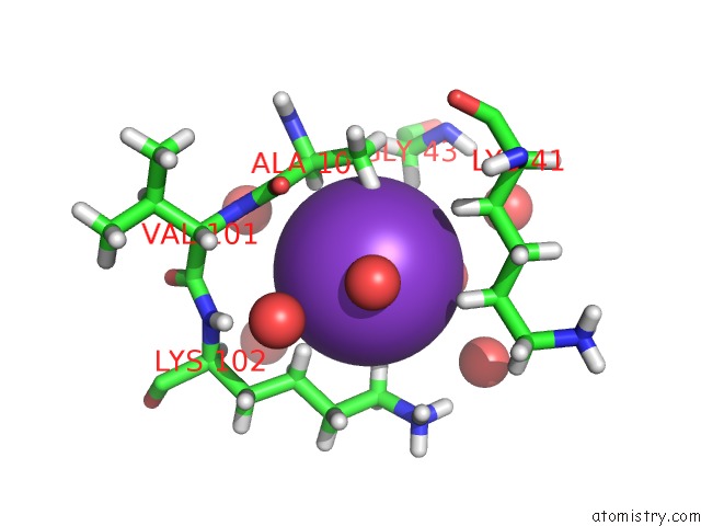
Mono view
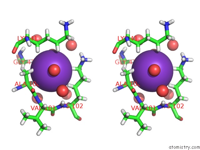
Stereo pair view
Mono view
Stereo pair view
A full contact list of Potassium with other atoms in the K binding
site number 1 of Crystal Structure of Human PAC3 Homodimer (Trigonal Form) within 5.0Å range:
|
Reference:
T.Satoh,
M.Yagi-Utsumi,
K.Okamoto,
E.Kurimoto,
K.Tanaka,
K.Kato.
Molecular and Structural Basis of the Proteasome Alpha Subunit Assembly Mechanism Mediated By the Proteasome-Assembling Chaperone PAC3-PAC4 Heterodimer. Int J Mol Sci V. 20 2019.
ISSN: ESSN 1422-0067
PubMed: 31067643
DOI: 10.3390/IJMS20092231
Page generated: Mon Aug 12 16:43:22 2024
ISSN: ESSN 1422-0067
PubMed: 31067643
DOI: 10.3390/IJMS20092231
Last articles
Zn in 9J0NZn in 9J0O
Zn in 9J0P
Zn in 9FJX
Zn in 9EKB
Zn in 9C0F
Zn in 9CAH
Zn in 9CH0
Zn in 9CH3
Zn in 9CH1