Potassium in PDB 6jjf: Crystal Structure of A Two-Quartet Dna Mixed-Parallel/Antiparallel G- Quadruplex
Protein crystallography data
The structure of Crystal Structure of A Two-Quartet Dna Mixed-Parallel/Antiparallel G- Quadruplex, PDB code: 6jjf
was solved by
Y.S.Zhang,
K.Ei Omari,
R.Duman,
A.Wagner,
G.N.Parkinson,
D.G.Wei,
with X-Ray Crystallography technique. A brief refinement statistics is given in the table below:
Resolution Low / High (Å)
|
23.80 /
1.47
|
Space group
|
C 1 2 1
|
Cell size a, b, c (Å), α, β, γ (°)
|
45.370,
47.600,
37.730,
90.00,
110.02,
90.00
|
R / Rfree (%)
|
19.2 /
19.9
|
Other elements in 6jjf:
The structure of Crystal Structure of A Two-Quartet Dna Mixed-Parallel/Antiparallel G- Quadruplex also contains other interesting chemical elements:
Potassium Binding Sites:
The binding sites of Potassium atom in the Crystal Structure of A Two-Quartet Dna Mixed-Parallel/Antiparallel G- Quadruplex
(pdb code 6jjf). This binding sites where shown within
5.0 Angstroms radius around Potassium atom.
In total 3 binding sites of Potassium where determined in the
Crystal Structure of A Two-Quartet Dna Mixed-Parallel/Antiparallel G- Quadruplex, PDB code: 6jjf:
Jump to Potassium binding site number:
1;
2;
3;
Potassium binding site 1 out
of 3 in 6jjf
Go back to
Potassium Binding Sites List in 6jjf
Potassium binding site 1 out
of 3 in the Crystal Structure of A Two-Quartet Dna Mixed-Parallel/Antiparallel G- Quadruplex
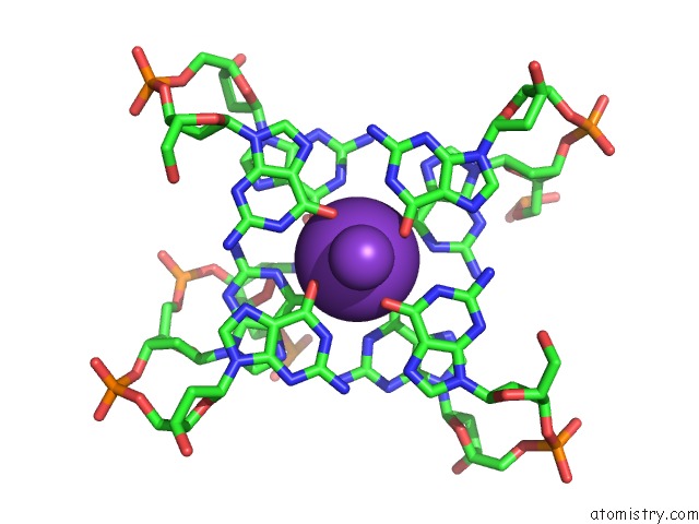
 Mono view
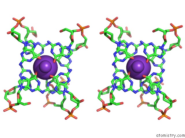
 Stereo pair view
|
A full contact list of Potassium with other atoms in the K binding
site number 1 of Crystal Structure of A Two-Quartet Dna Mixed-Parallel/Antiparallel G- Quadruplex within 5.0Å range:
probe
|
atom
|
residue
|
distance (Å)
|
B
|
Occ
|
A:K101
b:24.6
occ:1.00
|
O6
|
B:DG6
|
2.7
|
26.4
|
1.0
|
O6
|
A:DG6
|
2.7
|
26.1
|
1.0
|
O6
|
B:DG2
|
2.7
|
28.0
|
1.0
|
O6
|
A:DG2
|
2.8
|
25.8
|
1.0
|
O6
|
A:DG1
|
2.9
|
23.8
|
1.0
|
O6
|
B:DG1
|
2.9
|
22.9
|
1.0
|
O6
|
A:DG7
|
2.9
|
25.6
|
1.0
|
O6
|
B:DG7
|
2.9
|
25.1
|
1.0
|
K
|
A:K102
|
3.5
|
23.5
|
1.0
|
C6
|
A:DG6
|
3.6
|
24.0
|
1.0
|
C6
|
B:DG6
|
3.6
|
25.5
|
1.0
|
C6
|
B:DG2
|
3.7
|
25.6
|
1.0
|
C6
|
A:DG2
|
3.7
|
24.6
|
1.0
|
C6
|
A:DG1
|
3.7
|
23.2
|
1.0
|
C6
|
B:DG1
|
3.7
|
23.6
|
1.0
|
C6
|
A:DG7
|
3.8
|
24.1
|
1.0
|
C6
|
B:DG7
|
3.8
|
26.2
|
1.0
|
N1
|
B:DG6
|
3.8
|
26.8
|
1.0
|
N1
|
A:DG6
|
3.9
|
26.1
|
1.0
|
N1
|
B:DG2
|
3.9
|
27.0
|
1.0
|
N1
|
A:DG2
|
4.0
|
23.0
|
1.0
|
N1
|
A:DG7
|
4.0
|
22.7
|
1.0
|
N1
|
B:DG7
|
4.1
|
23.9
|
1.0
|
N1
|
B:DG1
|
4.1
|
23.1
|
1.0
|
N1
|
A:DG1
|
4.1
|
23.4
|
1.0
|
C4
|
B:DC5
|
4.5
|
26.5
|
1.0
|
N3
|
B:DC5
|
4.6
|
27.3
|
1.0
|
C5
|
B:DC5
|
4.6
|
30.8
|
1.0
|
N4
|
B:DC5
|
4.8
|
29.1
|
1.0
|
C5
|
A:DG6
|
4.8
|
25.5
|
1.0
|
C2
|
B:DC5
|
4.8
|
29.2
|
1.0
|
C6
|
B:DC5
|
4.9
|
28.2
|
1.0
|
C5
|
B:DG6
|
4.9
|
25.3
|
1.0
|
C5
|
A:DG1
|
4.9
|
24.2
|
1.0
|
C5
|
B:DG1
|
4.9
|
23.9
|
1.0
|
N1
|
B:DC5
|
5.0
|
26.1
|
1.0
|
C5
|
B:DG2
|
5.0
|
27.0
|
1.0
|
C5
|
A:DG7
|
5.0
|
24.1
|
1.0
|
|
Potassium binding site 2 out
of 3 in 6jjf
Go back to
Potassium Binding Sites List in 6jjf
Potassium binding site 2 out
of 3 in the Crystal Structure of A Two-Quartet Dna Mixed-Parallel/Antiparallel G- Quadruplex
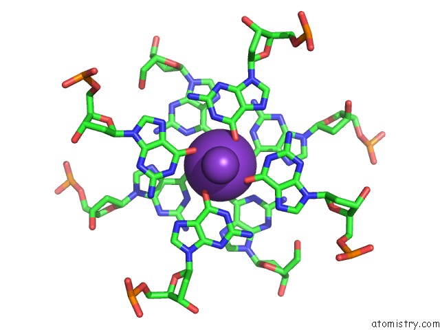
 Mono view
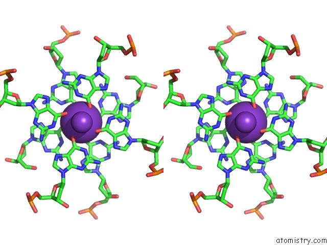
 Stereo pair view
|
A full contact list of Potassium with other atoms in the K binding
site number 2 of Crystal Structure of A Two-Quartet Dna Mixed-Parallel/Antiparallel G- Quadruplex within 5.0Å range:
probe
|
atom
|
residue
|
distance (Å)
|
B
|
Occ
|
A:K102
b:23.5
occ:1.00
|
O6
|
B:DG7
|
2.8
|
25.1
|
1.0
|
O6
|
B:DG12
|
2.8
|
21.6
|
1.0
|
O6
|
B:DG9
|
2.8
|
23.9
|
1.0
|
O6
|
A:DG12
|
2.8
|
22.7
|
1.0
|
O6
|
B:DG1
|
2.8
|
22.9
|
1.0
|
O6
|
A:DG9
|
2.8
|
21.5
|
1.0
|
O6
|
A:DG7
|
2.8
|
25.6
|
1.0
|
O6
|
A:DG1
|
2.8
|
23.8
|
1.0
|
K
|
A:K103
|
3.4
|
22.0
|
1.0
|
K
|
A:K101
|
3.5
|
24.6
|
1.0
|
C6
|
A:DG9
|
3.6
|
24.0
|
1.0
|
C6
|
B:DG7
|
3.7
|
26.2
|
1.0
|
C6
|
A:DG7
|
3.7
|
24.1
|
1.0
|
C6
|
B:DG9
|
3.7
|
25.4
|
1.0
|
C6
|
B:DG1
|
3.7
|
23.6
|
1.0
|
C6
|
B:DG12
|
3.7
|
24.1
|
1.0
|
C6
|
A:DG1
|
3.7
|
23.2
|
1.0
|
C6
|
A:DG12
|
3.7
|
24.2
|
1.0
|
N1
|
A:DG9
|
3.9
|
23.3
|
1.0
|
N1
|
B:DG9
|
4.0
|
23.3
|
1.0
|
N1
|
B:DG1
|
4.0
|
23.1
|
1.0
|
N1
|
A:DG1
|
4.0
|
23.4
|
1.0
|
N1
|
A:DG7
|
4.0
|
22.7
|
1.0
|
N1
|
B:DG12
|
4.0
|
21.2
|
1.0
|
N1
|
B:DG7
|
4.0
|
23.9
|
1.0
|
N1
|
A:DG12
|
4.0
|
21.6
|
1.0
|
C5
|
B:DG7
|
4.8
|
24.6
|
1.0
|
C5
|
A:DG7
|
4.8
|
24.1
|
1.0
|
C5
|
A:DG9
|
4.9
|
23.6
|
1.0
|
C5
|
B:DG9
|
4.9
|
24.1
|
1.0
|
C5
|
B:DG1
|
4.9
|
23.9
|
1.0
|
C5
|
B:DG12
|
4.9
|
25.0
|
1.0
|
C5
|
A:DG12
|
4.9
|
23.3
|
1.0
|
C5
|
A:DG1
|
5.0
|
24.2
|
1.0
|
|
Potassium binding site 3 out
of 3 in 6jjf
Go back to
Potassium Binding Sites List in 6jjf
Potassium binding site 3 out
of 3 in the Crystal Structure of A Two-Quartet Dna Mixed-Parallel/Antiparallel G- Quadruplex
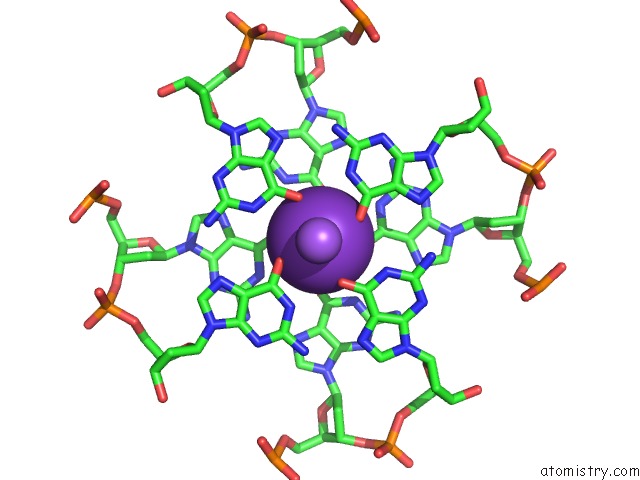
 Mono view
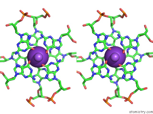
 Stereo pair view
|
A full contact list of Potassium with other atoms in the K binding
site number 3 of Crystal Structure of A Two-Quartet Dna Mixed-Parallel/Antiparallel G- Quadruplex within 5.0Å range:
probe
|
atom
|
residue
|
distance (Å)
|
B
|
Occ
|
A:K103
b:22.0
occ:1.00
|
O6
|
B:DG13
|
2.8
|
24.3
|
1.0
|
O6
|
A:DG13
|
2.8
|
23.1
|
1.0
|
O6
|
A:DG12
|
2.8
|
22.7
|
1.0
|
O6
|
B:DG12
|
2.9
|
21.6
|
1.0
|
O6
|
A:DG10
|
2.9
|
24.7
|
1.0
|
O6
|
B:DG10
|
2.9
|
22.6
|
1.0
|
O6
|
A:DG9
|
2.9
|
21.5
|
1.0
|
O6
|
B:DG9
|
2.9
|
23.9
|
1.0
|
K
|
A:K102
|
3.4
|
23.5
|
1.0
|
NA
|
A:NA106
|
3.5
|
27.9
|
1.0
|
C6
|
B:DG13
|
3.7
|
21.0
|
1.0
|
C6
|
A:DG13
|
3.7
|
22.0
|
1.0
|
C6
|
A:DG12
|
3.7
|
24.2
|
1.0
|
C6
|
B:DG12
|
3.7
|
24.1
|
1.0
|
C6
|
B:DG10
|
3.7
|
22.0
|
1.0
|
C6
|
A:DG10
|
3.7
|
24.8
|
1.0
|
C6
|
A:DG9
|
3.8
|
24.0
|
1.0
|
C6
|
B:DG9
|
3.8
|
25.4
|
1.0
|
N1
|
B:DG13
|
3.9
|
22.7
|
1.0
|
N1
|
A:DG13
|
4.0
|
21.9
|
1.0
|
N1
|
B:DG12
|
4.0
|
21.2
|
1.0
|
N1
|
B:DG10
|
4.0
|
22.5
|
1.0
|
N1
|
A:DG12
|
4.0
|
21.6
|
1.0
|
N1
|
A:DG10
|
4.0
|
24.3
|
1.0
|
N1
|
B:DG9
|
4.1
|
23.3
|
1.0
|
N1
|
A:DG9
|
4.1
|
23.3
|
1.0
|
C5
|
B:DG10
|
4.9
|
21.9
|
1.0
|
C5
|
A:DG10
|
4.9
|
22.6
|
1.0
|
C5
|
B:DG13
|
4.9
|
20.0
|
1.0
|
C5
|
A:DG12
|
4.9
|
23.3
|
1.0
|
C5
|
B:DG12
|
4.9
|
25.0
|
1.0
|
C5
|
A:DG13
|
4.9
|
22.7
|
1.0
|
|
Reference:
Y.S.Zhang,
K.Ei Omari,
R.Duman,
A.Wagner,
G.N.Parkinson,
D.G.Wei.
Crystal Structure of A Two-Quartet Dna Mixed-Parallel/Antiparallel G-Quadruplex To Be Published.
Page generated: Mon Aug 12 16:41:52 2024
|