Potassium »
PDB 6hae-6i4g »
6hrr »
Potassium in PDB 6hrr: Structure of the TRPML2 Eld at pH 6.5
Protein crystallography data
The structure of Structure of the TRPML2 Eld at pH 6.5, PDB code: 6hrr
was solved by
N.Bader,
K.K.Viet,
A.Wagner,
U.A.Hellmich,
with X-Ray Crystallography technique. A brief refinement statistics is given in the table below:
| Resolution Low / High (Å) | 20.00 / 2.00 |
| Space group | I 4 2 2 |
| Cell size a, b, c (Å), α, β, γ (°) | 109.471, 109.471, 149.744, 90.00, 90.00, 90.00 |
| R / Rfree (%) | 18.6 / 21.7 |
Other elements in 6hrr:
The structure of Structure of the TRPML2 Eld at pH 6.5 also contains other interesting chemical elements:
| Chlorine | (Cl) | 3 atoms |
Potassium Binding Sites:
The binding sites of Potassium atom in the Structure of the TRPML2 Eld at pH 6.5
(pdb code 6hrr). This binding sites where shown within
5.0 Angstroms radius around Potassium atom.
In total only one binding site of Potassium was determined in the Structure of the TRPML2 Eld at pH 6.5, PDB code: 6hrr:
In total only one binding site of Potassium was determined in the Structure of the TRPML2 Eld at pH 6.5, PDB code: 6hrr:
Potassium binding site 1 out of 1 in 6hrr
Go back to
Potassium binding site 1 out
of 1 in the Structure of the TRPML2 Eld at pH 6.5
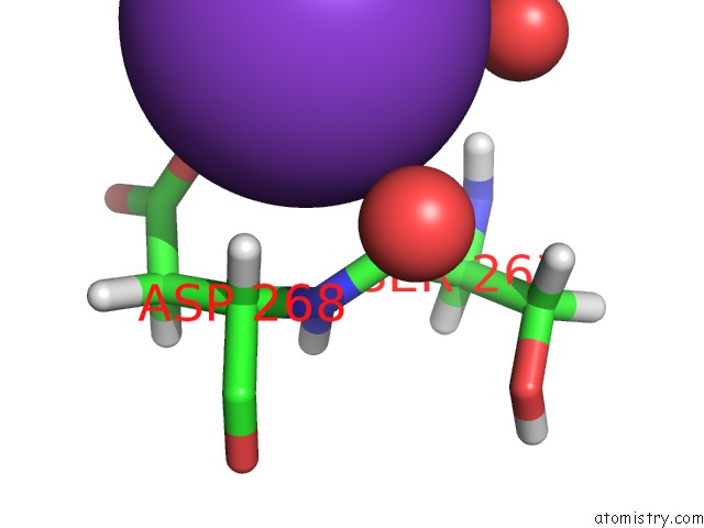
Mono view
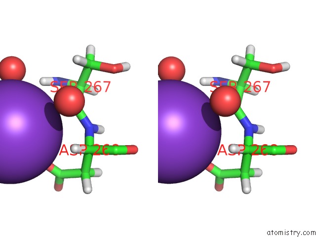
Stereo pair view
Mono view
Stereo pair view
A full contact list of Potassium with other atoms in the K binding
site number 1 of Structure of the TRPML2 Eld at pH 6.5 within 5.0Å range:
|
Reference:
K.K.Viet,
A.Wagner,
K.Schwickert,
N.Hellwig,
M.Brennich,
N.Bader,
T.Schirmeister,
N.Morgner,
H.Schindelin,
U.A.Hellmich.
Structure of the Human TRPML2 Ion Channel Extracytosolic/Lumenal Domain. Structure V. 27 1246 2019.
ISSN: ISSN 0969-2126
PubMed: 31178222
DOI: 10.1016/J.STR.2019.04.016
Page generated: Sat Aug 9 11:11:46 2025
ISSN: ISSN 0969-2126
PubMed: 31178222
DOI: 10.1016/J.STR.2019.04.016
Last articles
Mg in 3FXIMg in 3FWS
Mg in 3FWY
Mg in 3FWZ
Mg in 3FWA
Mg in 3FW9
Mg in 3FW8
Mg in 3FVB
Mg in 3FW7
Mg in 3FVY