Potassium »
PDB 6f3o-6h6x »
6h6x »
Potassium in PDB 6h6x: Structure of An Evolved Dimeric Form of the Ubid-Class Enzyme Hmff From Pelotomaculum Thermopropionicum in Complex with Prfmn
Protein crystallography data
The structure of Structure of An Evolved Dimeric Form of the Ubid-Class Enzyme Hmff From Pelotomaculum Thermopropionicum in Complex with Prfmn, PDB code: 6h6x
was solved by
K.A.P.Payne,
D.Leys,
with X-Ray Crystallography technique. A brief refinement statistics is given in the table below:
| Resolution Low / High (Å) | 83.86 / 2.25 |
| Space group | P 21 21 21 |
| Cell size a, b, c (Å), α, β, γ (°) | 167.720, 63.930, 98.300, 90.00, 90.00, 90.00 |
| R / Rfree (%) | 19.4 / 24.8 |
Other elements in 6h6x:
The structure of Structure of An Evolved Dimeric Form of the Ubid-Class Enzyme Hmff From Pelotomaculum Thermopropionicum in Complex with Prfmn also contains other interesting chemical elements:
| Manganese | (Mn) | 2 atoms |
| Calcium | (Ca) | 2 atoms |
Potassium Binding Sites:
The binding sites of Potassium atom in the Structure of An Evolved Dimeric Form of the Ubid-Class Enzyme Hmff From Pelotomaculum Thermopropionicum in Complex with Prfmn
(pdb code 6h6x). This binding sites where shown within
5.0 Angstroms radius around Potassium atom.
In total 2 binding sites of Potassium where determined in the Structure of An Evolved Dimeric Form of the Ubid-Class Enzyme Hmff From Pelotomaculum Thermopropionicum in Complex with Prfmn, PDB code: 6h6x:
Jump to Potassium binding site number: 1; 2;
In total 2 binding sites of Potassium where determined in the Structure of An Evolved Dimeric Form of the Ubid-Class Enzyme Hmff From Pelotomaculum Thermopropionicum in Complex with Prfmn, PDB code: 6h6x:
Jump to Potassium binding site number: 1; 2;
Potassium binding site 1 out of 2 in 6h6x
Go back to
Potassium binding site 1 out
of 2 in the Structure of An Evolved Dimeric Form of the Ubid-Class Enzyme Hmff From Pelotomaculum Thermopropionicum in Complex with Prfmn
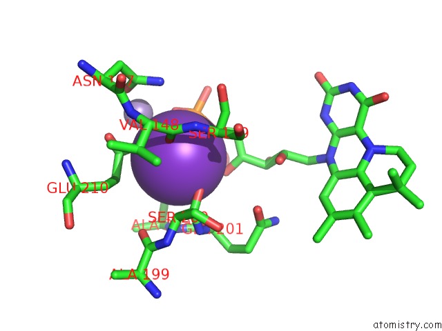
Mono view
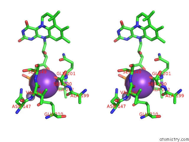
Stereo pair view
Mono view
Stereo pair view
A full contact list of Potassium with other atoms in the K binding
site number 1 of Structure of An Evolved Dimeric Form of the Ubid-Class Enzyme Hmff From Pelotomaculum Thermopropionicum in Complex with Prfmn within 5.0Å range:
|
Potassium binding site 2 out of 2 in 6h6x
Go back to
Potassium binding site 2 out
of 2 in the Structure of An Evolved Dimeric Form of the Ubid-Class Enzyme Hmff From Pelotomaculum Thermopropionicum in Complex with Prfmn
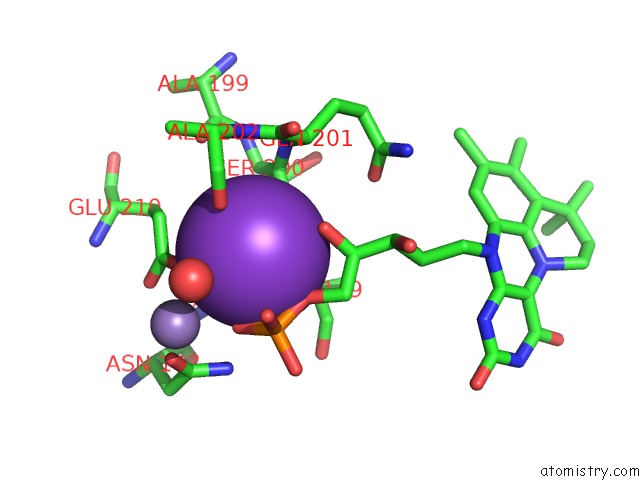
Mono view
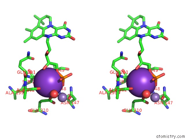
Stereo pair view
Mono view
Stereo pair view
A full contact list of Potassium with other atoms in the K binding
site number 2 of Structure of An Evolved Dimeric Form of the Ubid-Class Enzyme Hmff From Pelotomaculum Thermopropionicum in Complex with Prfmn within 5.0Å range:
|
Reference:
K.A.P.Payne,
S.A.Marshall,
K.Fisher,
M.J.Cliff,
D.M.Cannas,
C.Yan,
D.J.Heyes,
D.A.Parker,
I.Larrosa,
D.Leys.
Enzymatic Carboxylation of 2-Furoic Acid Yields 2,5-Furandicarboxylic Acid (Fdca). Acs Catalysis V. 9 2854 2019.
ISSN: ESSN 2155-5435
PubMed: 31057985
DOI: 10.1021/ACSCATAL.8B04862
Page generated: Mon Aug 12 16:20:55 2024
ISSN: ESSN 2155-5435
PubMed: 31057985
DOI: 10.1021/ACSCATAL.8B04862
Last articles
Zn in 9J0NZn in 9J0O
Zn in 9J0P
Zn in 9FJX
Zn in 9EKB
Zn in 9C0F
Zn in 9CAH
Zn in 9CH0
Zn in 9CH3
Zn in 9CH1