Potassium »
PDB 6f3o-6h6x »
6gx3 »
Potassium in PDB 6gx3: Crystal Structure of Schistosoma Mansoni HDAC8 Complexed with An Hydroxamate 1
Enzymatic activity of Crystal Structure of Schistosoma Mansoni HDAC8 Complexed with An Hydroxamate 1
All present enzymatic activity of Crystal Structure of Schistosoma Mansoni HDAC8 Complexed with An Hydroxamate 1:
3.5.1.98;
3.5.1.98;
Protein crystallography data
The structure of Crystal Structure of Schistosoma Mansoni HDAC8 Complexed with An Hydroxamate 1, PDB code: 6gx3
was solved by
T.B.Shaik,
M.Marek,
C.Romier,
with X-Ray Crystallography technique. A brief refinement statistics is given in the table below:
| Resolution Low / High (Å) | 47.89 / 2.10 |
| Space group | P 1 |
| Cell size a, b, c (Å), α, β, γ (°) | 70.449, 70.670, 98.413, 75.71, 78.40, 85.55 |
| R / Rfree (%) | 18.9 / 24 |
Other elements in 6gx3:
The structure of Crystal Structure of Schistosoma Mansoni HDAC8 Complexed with An Hydroxamate 1 also contains other interesting chemical elements:
| Chlorine | (Cl) | 8 atoms |
| Zinc | (Zn) | 4 atoms |
Potassium Binding Sites:
The binding sites of Potassium atom in the Crystal Structure of Schistosoma Mansoni HDAC8 Complexed with An Hydroxamate 1
(pdb code 6gx3). This binding sites where shown within
5.0 Angstroms radius around Potassium atom.
In total 8 binding sites of Potassium where determined in the Crystal Structure of Schistosoma Mansoni HDAC8 Complexed with An Hydroxamate 1, PDB code: 6gx3:
Jump to Potassium binding site number: 1; 2; 3; 4; 5; 6; 7; 8;
In total 8 binding sites of Potassium where determined in the Crystal Structure of Schistosoma Mansoni HDAC8 Complexed with An Hydroxamate 1, PDB code: 6gx3:
Jump to Potassium binding site number: 1; 2; 3; 4; 5; 6; 7; 8;
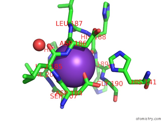
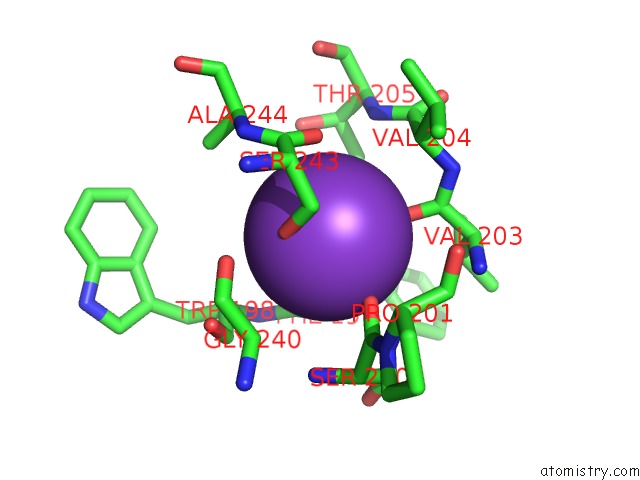
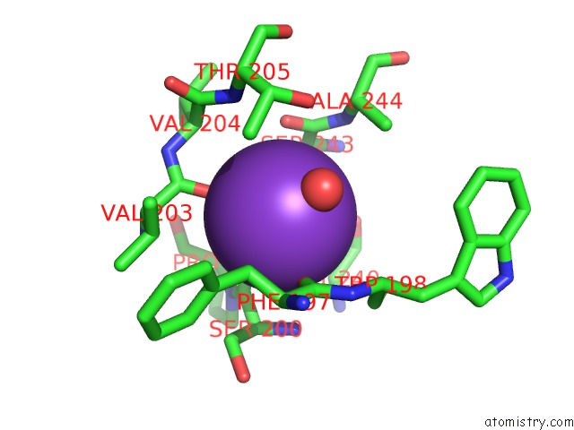
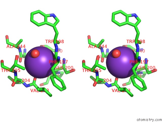
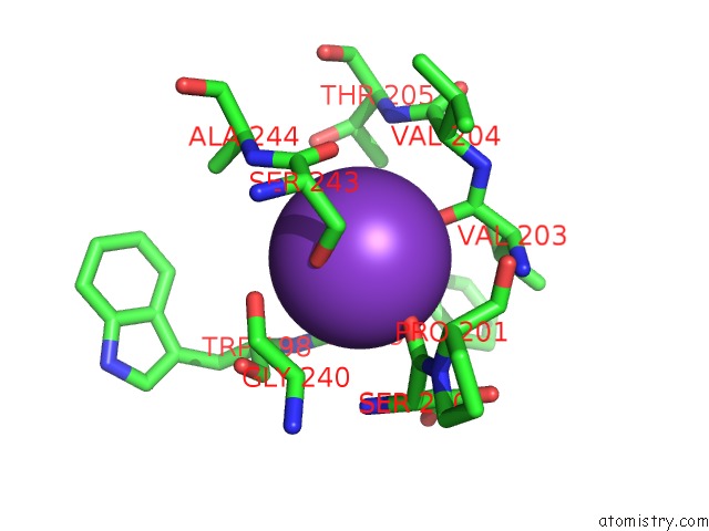
Potassium binding site 1 out of 8 in 6gx3
Go back to
Potassium binding site 1 out
of 8 in the Crystal Structure of Schistosoma Mansoni HDAC8 Complexed with An Hydroxamate 1
Mono view
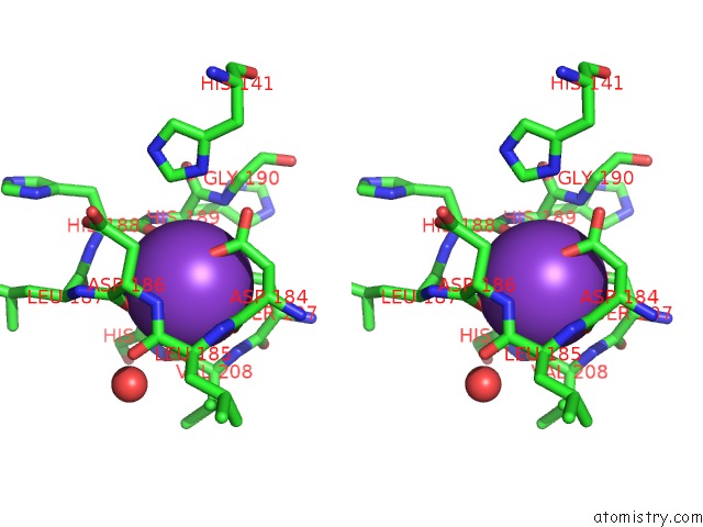
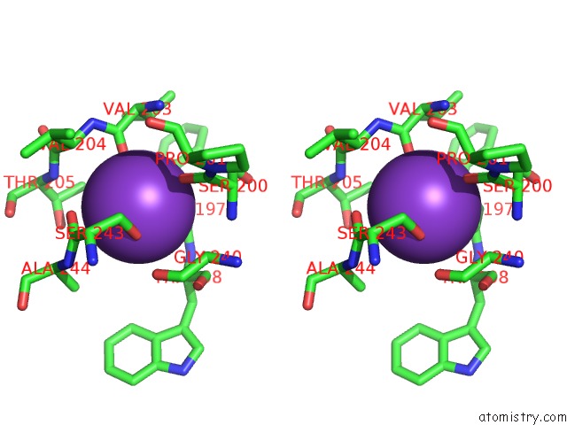
Stereo pair view
Mono view
Stereo pair view
A full contact list of Potassium with other atoms in the K binding
site number 1 of Crystal Structure of Schistosoma Mansoni HDAC8 Complexed with An Hydroxamate 1 within 5.0Å range:
|
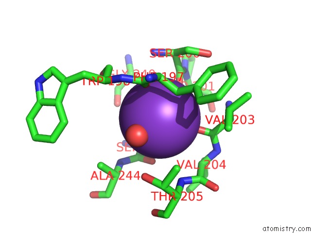
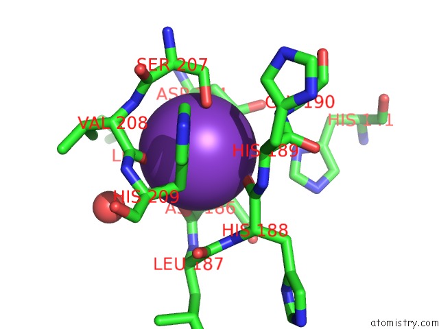
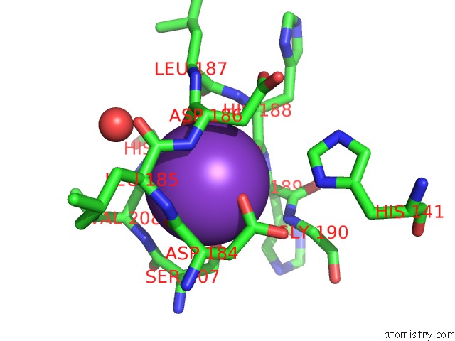
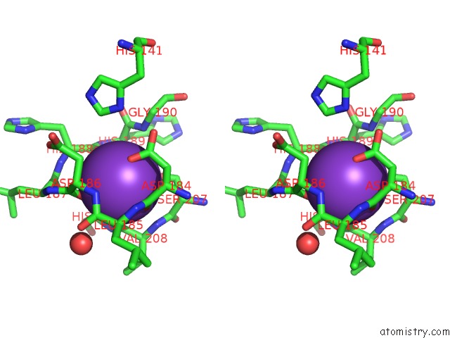
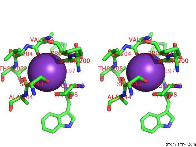
Potassium binding site 2 out of 8 in 6gx3
Go back to
Potassium binding site 2 out
of 8 in the Crystal Structure of Schistosoma Mansoni HDAC8 Complexed with An Hydroxamate 1
Mono view
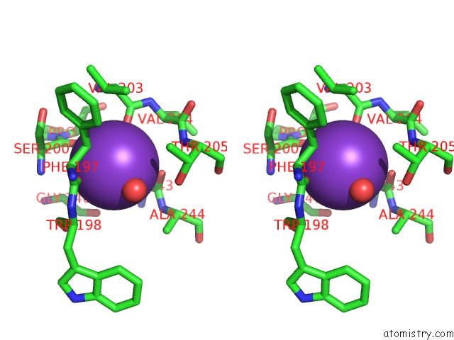
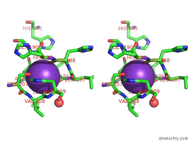
Stereo pair view
Mono view
Stereo pair view
A full contact list of Potassium with other atoms in the K binding
site number 2 of Crystal Structure of Schistosoma Mansoni HDAC8 Complexed with An Hydroxamate 1 within 5.0Å range:
|
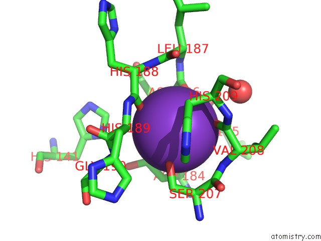
Potassium binding site 3 out of 8 in 6gx3
Go back to
Potassium binding site 3 out
of 8 in the Crystal Structure of Schistosoma Mansoni HDAC8 Complexed with An Hydroxamate 1
Mono view
Stereo pair view
Mono view
Stereo pair view
A full contact list of Potassium with other atoms in the K binding
site number 3 of Crystal Structure of Schistosoma Mansoni HDAC8 Complexed with An Hydroxamate 1 within 5.0Å range:
|
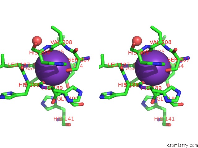
Potassium binding site 4 out of 8 in 6gx3
Go back to
Potassium binding site 4 out
of 8 in the Crystal Structure of Schistosoma Mansoni HDAC8 Complexed with An Hydroxamate 1
Mono view
Stereo pair view
Mono view
Stereo pair view
A full contact list of Potassium with other atoms in the K binding
site number 4 of Crystal Structure of Schistosoma Mansoni HDAC8 Complexed with An Hydroxamate 1 within 5.0Å range:
|
Potassium binding site 5 out of 8 in 6gx3
Go back to
Potassium binding site 5 out
of 8 in the Crystal Structure of Schistosoma Mansoni HDAC8 Complexed with An Hydroxamate 1
Mono view
Stereo pair view
Mono view
Stereo pair view
A full contact list of Potassium with other atoms in the K binding
site number 5 of Crystal Structure of Schistosoma Mansoni HDAC8 Complexed with An Hydroxamate 1 within 5.0Å range:
|
Potassium binding site 6 out of 8 in 6gx3
Go back to
Potassium binding site 6 out
of 8 in the Crystal Structure of Schistosoma Mansoni HDAC8 Complexed with An Hydroxamate 1
Mono view
Stereo pair view
Mono view
Stereo pair view
A full contact list of Potassium with other atoms in the K binding
site number 6 of Crystal Structure of Schistosoma Mansoni HDAC8 Complexed with An Hydroxamate 1 within 5.0Å range:
|
Potassium binding site 7 out of 8 in 6gx3
Go back to
Potassium binding site 7 out
of 8 in the Crystal Structure of Schistosoma Mansoni HDAC8 Complexed with An Hydroxamate 1
Mono view
Stereo pair view
Mono view
Stereo pair view
A full contact list of Potassium with other atoms in the K binding
site number 7 of Crystal Structure of Schistosoma Mansoni HDAC8 Complexed with An Hydroxamate 1 within 5.0Å range:
|
Potassium binding site 8 out of 8 in 6gx3
Go back to
Potassium binding site 8 out
of 8 in the Crystal Structure of Schistosoma Mansoni HDAC8 Complexed with An Hydroxamate 1
Mono view
Stereo pair view
Mono view
Stereo pair view
A full contact list of Potassium with other atoms in the K binding
site number 8 of Crystal Structure of Schistosoma Mansoni HDAC8 Complexed with An Hydroxamate 1 within 5.0Å range:
|
Reference:
T.Bayer,
A.Chakrabarti,
J.Lancelot,
T.B.Shaik,
K.Hausmann,
J.Melesina,
K.Schmidtkunz,
M.Marek,
F.Erdmann,
M.Schmidt,
D.Robaa,
C.Romier,
R.J.Pierce,
M.Jung,
W.Sippl.
Synthesis, Crystallization Studies, and in Vitro Characterization of Cinnamic Acid Derivatives As SMHDAC8 Inhibitors For the Treatment of Schistosomiasis. Chemmedchem V. 13 1517 2018.
ISSN: ESSN 1860-7187
PubMed: 29806110
DOI: 10.1002/CMDC.201800238
Page generated: Mon Aug 12 16:14:51 2024
ISSN: ESSN 1860-7187
PubMed: 29806110
DOI: 10.1002/CMDC.201800238
Last articles
Zn in 9J0NZn in 9J0O
Zn in 9J0P
Zn in 9FJX
Zn in 9EKB
Zn in 9C0F
Zn in 9CAH
Zn in 9CH0
Zn in 9CH3
Zn in 9CH1