Potassium »
PDB 6f3o-6h6x »
6gg5 »
Potassium in PDB 6gg5: Crystal Structure of M2 Pyk in Complex with Tryptophan.
Enzymatic activity of Crystal Structure of M2 Pyk in Complex with Tryptophan.
All present enzymatic activity of Crystal Structure of M2 Pyk in Complex with Tryptophan.:
2.7.1.40;
2.7.1.40;
Protein crystallography data
The structure of Crystal Structure of M2 Pyk in Complex with Tryptophan., PDB code: 6gg5
was solved by
I.W.Mcnae,
M.Yuan,
M.D.Walkinshaw,
with X-Ray Crystallography technique. A brief refinement statistics is given in the table below:
| Resolution Low / High (Å) | 72.54 / 3.20 |
| Space group | P 1 21 1 |
| Cell size a, b, c (Å), α, β, γ (°) | 96.880, 70.576, 168.502, 90.00, 106.02, 90.00 |
| R / Rfree (%) | 22.8 / 26.5 |
Potassium Binding Sites:
The binding sites of Potassium atom in the Crystal Structure of M2 Pyk in Complex with Tryptophan.
(pdb code 6gg5). This binding sites where shown within
5.0 Angstroms radius around Potassium atom.
In total 4 binding sites of Potassium where determined in the Crystal Structure of M2 Pyk in Complex with Tryptophan., PDB code: 6gg5:
Jump to Potassium binding site number: 1; 2; 3; 4;
In total 4 binding sites of Potassium where determined in the Crystal Structure of M2 Pyk in Complex with Tryptophan., PDB code: 6gg5:
Jump to Potassium binding site number: 1; 2; 3; 4;
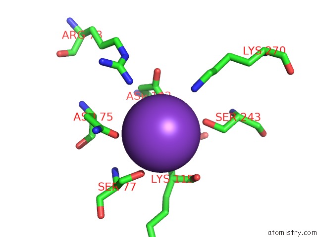
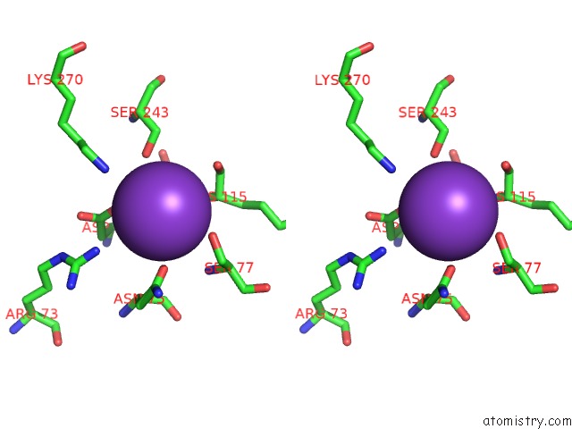
Potassium binding site 1 out of 4 in 6gg5
Go back to
Potassium binding site 1 out
of 4 in the Crystal Structure of M2 Pyk in Complex with Tryptophan.
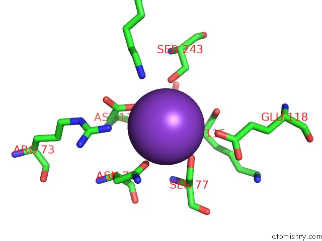
Mono view
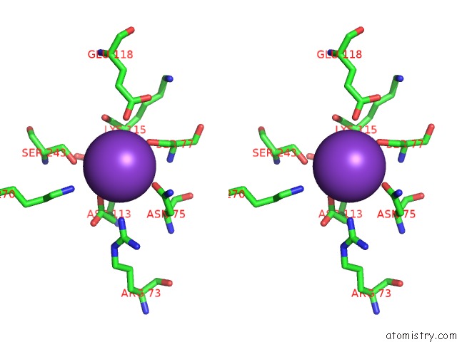
Stereo pair view
Mono view
Stereo pair view
A full contact list of Potassium with other atoms in the K binding
site number 1 of Crystal Structure of M2 Pyk in Complex with Tryptophan. within 5.0Å range:
|
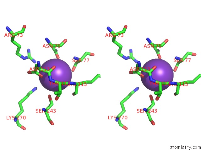
Potassium binding site 2 out of 4 in 6gg5
Go back to
Potassium binding site 2 out
of 4 in the Crystal Structure of M2 Pyk in Complex with Tryptophan.
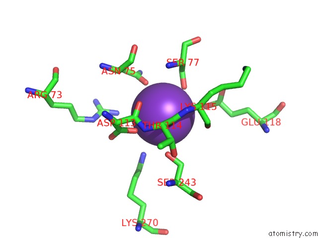
Mono view
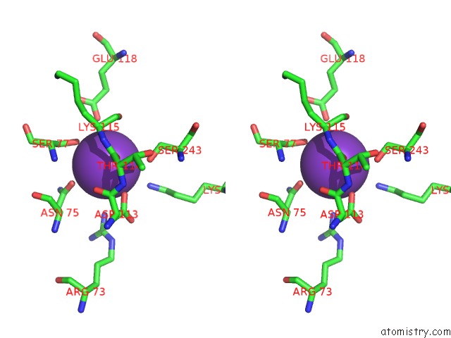
Stereo pair view
Mono view
Stereo pair view
A full contact list of Potassium with other atoms in the K binding
site number 2 of Crystal Structure of M2 Pyk in Complex with Tryptophan. within 5.0Å range:
|
Potassium binding site 3 out of 4 in 6gg5
Go back to
Potassium binding site 3 out
of 4 in the Crystal Structure of M2 Pyk in Complex with Tryptophan.
Mono view
Stereo pair view
Mono view
Stereo pair view
A full contact list of Potassium with other atoms in the K binding
site number 3 of Crystal Structure of M2 Pyk in Complex with Tryptophan. within 5.0Å range:
|
Potassium binding site 4 out of 4 in 6gg5
Go back to
Potassium binding site 4 out
of 4 in the Crystal Structure of M2 Pyk in Complex with Tryptophan.

Mono view
Stereo pair view
Mono view
Stereo pair view
A full contact list of Potassium with other atoms in the K binding
site number 4 of Crystal Structure of M2 Pyk in Complex with Tryptophan. within 5.0Å range:
|
Reference:
M.Yuan,
I.W.Mcnae,
Y.Chen,
E.A.Blackburn,
M.A.Wear,
P.A.M.Michels,
L.A.Fothergill-Gilmore,
T.Hupp,
M.D.Walkinshaw.
An Allostatic Mechanism For M2 Pyruvate Kinase As An Amino-Acid Sensor. Biochem. J. V. 475 1821 2018.
ISSN: ESSN 1470-8728
PubMed: 29748232
DOI: 10.1042/BCJ20180171
Page generated: Mon Aug 12 16:09:17 2024
ISSN: ESSN 1470-8728
PubMed: 29748232
DOI: 10.1042/BCJ20180171
Last articles
Zn in 9JYWZn in 9IR4
Zn in 9IR3
Zn in 9GMX
Zn in 9GMW
Zn in 9JEJ
Zn in 9ERF
Zn in 9ERE
Zn in 9EGV
Zn in 9EGW