Potassium »
PDB 6f3o-6h6x »
6f3o »
Potassium in PDB 6f3o: Crystal Structure of S-Adenosyl-L-Homocysteine Hydrolase From Pseudomonas Aeruginosa Complexed with Adenine, K+ and ZN2+ Cations
Enzymatic activity of Crystal Structure of S-Adenosyl-L-Homocysteine Hydrolase From Pseudomonas Aeruginosa Complexed with Adenine, K+ and ZN2+ Cations
All present enzymatic activity of Crystal Structure of S-Adenosyl-L-Homocysteine Hydrolase From Pseudomonas Aeruginosa Complexed with Adenine, K+ and ZN2+ Cations:
3.3.1.1;
3.3.1.1;
Protein crystallography data
The structure of Crystal Structure of S-Adenosyl-L-Homocysteine Hydrolase From Pseudomonas Aeruginosa Complexed with Adenine, K+ and ZN2+ Cations, PDB code: 6f3o
was solved by
J.Czyrko,
K.Brzezinski,
with X-Ray Crystallography technique. A brief refinement statistics is given in the table below:
| Resolution Low / High (Å) | 25.00 / 1.75 |
| Space group | C 1 2 1 |
| Cell size a, b, c (Å), α, β, γ (°) | 170.839, 99.614, 111.825, 90.00, 101.97, 90.00 |
| R / Rfree (%) | 14 / 16.7 |
Other elements in 6f3o:
The structure of Crystal Structure of S-Adenosyl-L-Homocysteine Hydrolase From Pseudomonas Aeruginosa Complexed with Adenine, K+ and ZN2+ Cations also contains other interesting chemical elements:
| Zinc | (Zn) | 2 atoms |
Potassium Binding Sites:
The binding sites of Potassium atom in the Crystal Structure of S-Adenosyl-L-Homocysteine Hydrolase From Pseudomonas Aeruginosa Complexed with Adenine, K+ and ZN2+ Cations
(pdb code 6f3o). This binding sites where shown within
5.0 Angstroms radius around Potassium atom.
In total 4 binding sites of Potassium where determined in the Crystal Structure of S-Adenosyl-L-Homocysteine Hydrolase From Pseudomonas Aeruginosa Complexed with Adenine, K+ and ZN2+ Cations, PDB code: 6f3o:
Jump to Potassium binding site number: 1; 2; 3; 4;
In total 4 binding sites of Potassium where determined in the Crystal Structure of S-Adenosyl-L-Homocysteine Hydrolase From Pseudomonas Aeruginosa Complexed with Adenine, K+ and ZN2+ Cations, PDB code: 6f3o:
Jump to Potassium binding site number: 1; 2; 3; 4;
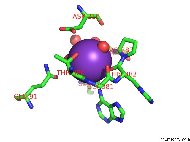
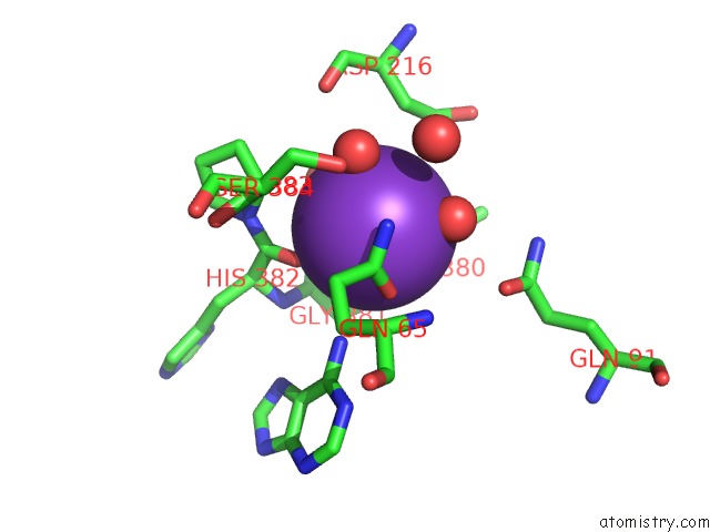
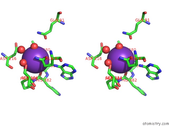
Potassium binding site 1 out of 4 in 6f3o
Go back to
Potassium binding site 1 out
of 4 in the Crystal Structure of S-Adenosyl-L-Homocysteine Hydrolase From Pseudomonas Aeruginosa Complexed with Adenine, K+ and ZN2+ Cations
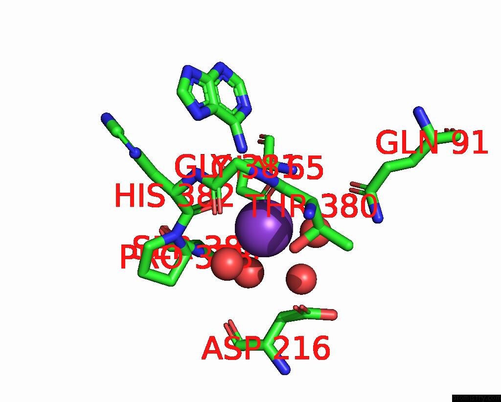
Mono view
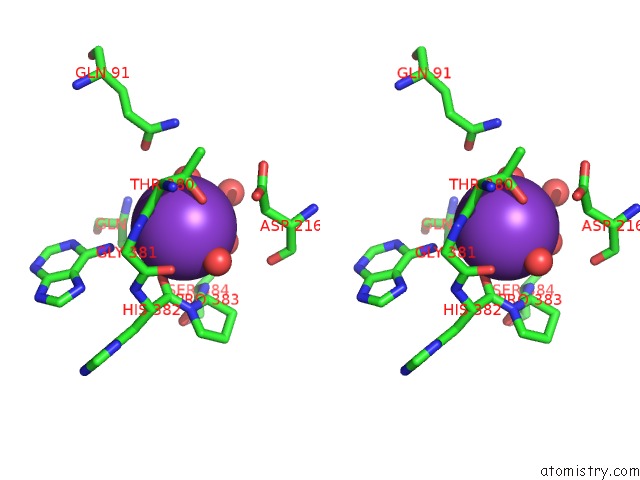
Stereo pair view
Mono view
Stereo pair view
A full contact list of Potassium with other atoms in the K binding
site number 1 of Crystal Structure of S-Adenosyl-L-Homocysteine Hydrolase From Pseudomonas Aeruginosa Complexed with Adenine, K+ and ZN2+ Cations within 5.0Å range:
|
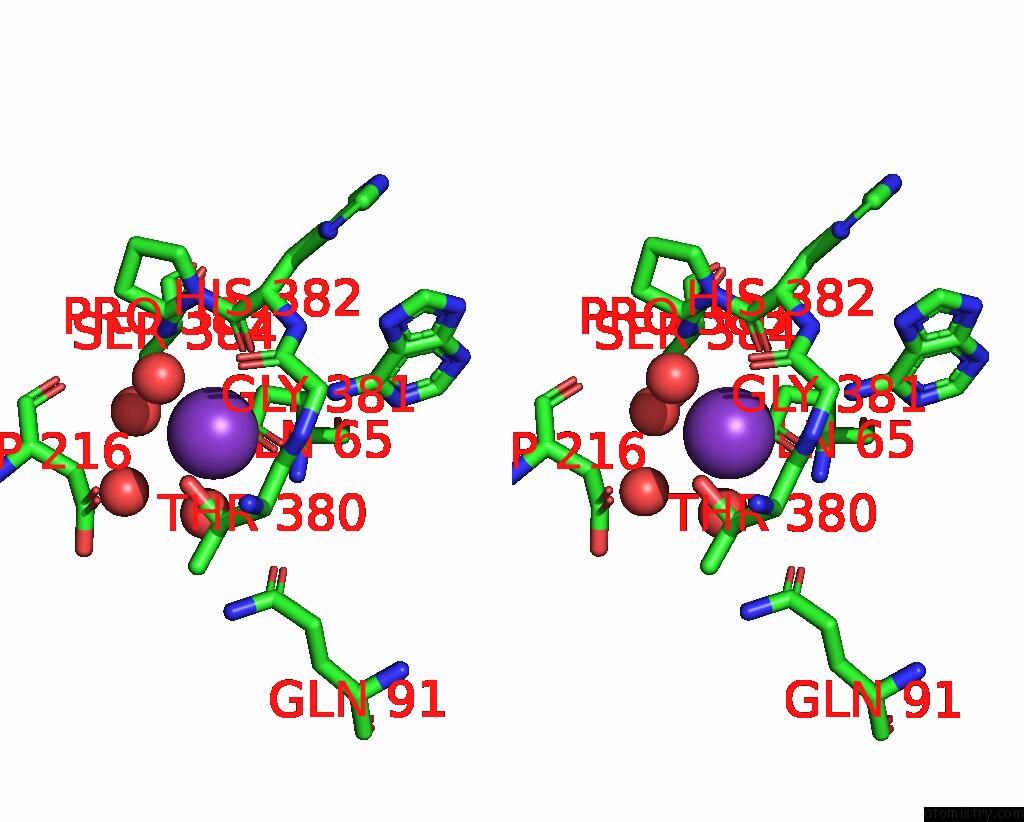
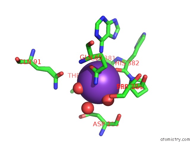
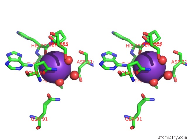
Potassium binding site 2 out of 4 in 6f3o
Go back to
Potassium binding site 2 out
of 4 in the Crystal Structure of S-Adenosyl-L-Homocysteine Hydrolase From Pseudomonas Aeruginosa Complexed with Adenine, K+ and ZN2+ Cations
Mono view
Stereo pair view
Mono view
Stereo pair view
A full contact list of Potassium with other atoms in the K binding
site number 2 of Crystal Structure of S-Adenosyl-L-Homocysteine Hydrolase From Pseudomonas Aeruginosa Complexed with Adenine, K+ and ZN2+ Cations within 5.0Å range:
|
Potassium binding site 3 out of 4 in 6f3o
Go back to
Potassium binding site 3 out
of 4 in the Crystal Structure of S-Adenosyl-L-Homocysteine Hydrolase From Pseudomonas Aeruginosa Complexed with Adenine, K+ and ZN2+ Cations
Mono view
Stereo pair view
Mono view
Stereo pair view
A full contact list of Potassium with other atoms in the K binding
site number 3 of Crystal Structure of S-Adenosyl-L-Homocysteine Hydrolase From Pseudomonas Aeruginosa Complexed with Adenine, K+ and ZN2+ Cations within 5.0Å range:
|
Potassium binding site 4 out of 4 in 6f3o
Go back to
Potassium binding site 4 out
of 4 in the Crystal Structure of S-Adenosyl-L-Homocysteine Hydrolase From Pseudomonas Aeruginosa Complexed with Adenine, K+ and ZN2+ Cations
Mono view
Stereo pair view
Mono view
Stereo pair view
A full contact list of Potassium with other atoms in the K binding
site number 4 of Crystal Structure of S-Adenosyl-L-Homocysteine Hydrolase From Pseudomonas Aeruginosa Complexed with Adenine, K+ and ZN2+ Cations within 5.0Å range:
|
Reference:
J.Czyrko,
J.Sliwiak,
B.Imiolczyk,
Z.Gdaniec,
M.Jaskolski,
K.Brzezinski.
Metal-Cation Regulation of Enzyme Dynamics Is A Key Factor Influencing the Activity of S-Adenosyl-L-Homocysteine Hydrolase From Pseudomonas Aeruginosa. Sci Rep V. 8 11334 2018.
ISSN: ESSN 2045-2322
PubMed: 30054521
DOI: 10.1038/S41598-018-29535-Y
Page generated: Mon Aug 12 16:06:05 2024
ISSN: ESSN 2045-2322
PubMed: 30054521
DOI: 10.1038/S41598-018-29535-Y
Last articles
Zn in 9J0NZn in 9J0O
Zn in 9J0P
Zn in 9FJX
Zn in 9EKB
Zn in 9C0F
Zn in 9CAH
Zn in 9CH0
Zn in 9CH3
Zn in 9CH1