Potassium »
PDB 6dxz-6f3n »
6eng »
Potassium in PDB 6eng: Crystal Structure of the 43K Atpase Domain of Escherichia Coli Gyrase B in Complex with An Aminocoumarin
Enzymatic activity of Crystal Structure of the 43K Atpase Domain of Escherichia Coli Gyrase B in Complex with An Aminocoumarin
All present enzymatic activity of Crystal Structure of the 43K Atpase Domain of Escherichia Coli Gyrase B in Complex with An Aminocoumarin:
5.99.1.3;
5.99.1.3;
Protein crystallography data
The structure of Crystal Structure of the 43K Atpase Domain of Escherichia Coli Gyrase B in Complex with An Aminocoumarin, PDB code: 6eng
was solved by
A.Vanden Broeck,
A.G.Mcewen,
V.Lamour,
with X-Ray Crystallography technique. A brief refinement statistics is given in the table below:
| Resolution Low / High (Å) | 33.67 / 2.30 |
| Space group | P 21 21 21 |
| Cell size a, b, c (Å), α, β, γ (°) | 47.830, 128.070, 159.760, 90.00, 90.00, 90.00 |
| R / Rfree (%) | 20 / 25.7 |
Other elements in 6eng:
The structure of Crystal Structure of the 43K Atpase Domain of Escherichia Coli Gyrase B in Complex with An Aminocoumarin also contains other interesting chemical elements:
| Chlorine | (Cl) | 2 atoms |
| Sodium | (Na) | 1 atom |
Potassium Binding Sites:
The binding sites of Potassium atom in the Crystal Structure of the 43K Atpase Domain of Escherichia Coli Gyrase B in Complex with An Aminocoumarin
(pdb code 6eng). This binding sites where shown within
5.0 Angstroms radius around Potassium atom.
In total 3 binding sites of Potassium where determined in the Crystal Structure of the 43K Atpase Domain of Escherichia Coli Gyrase B in Complex with An Aminocoumarin, PDB code: 6eng:
Jump to Potassium binding site number: 1; 2; 3;
In total 3 binding sites of Potassium where determined in the Crystal Structure of the 43K Atpase Domain of Escherichia Coli Gyrase B in Complex with An Aminocoumarin, PDB code: 6eng:
Jump to Potassium binding site number: 1; 2; 3;
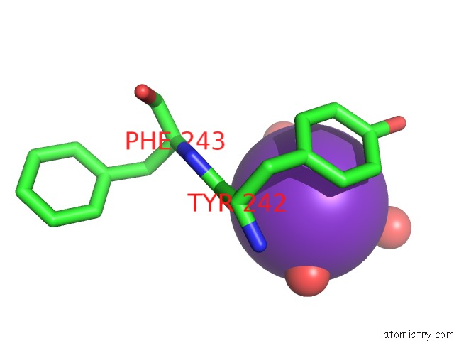
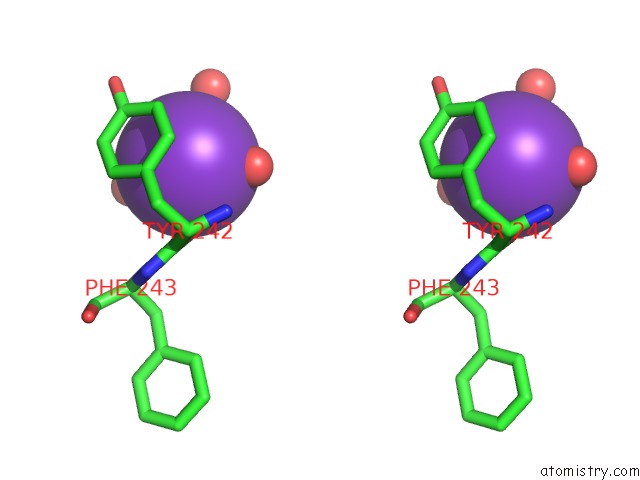
Potassium binding site 1 out of 3 in 6eng
Go back to
Potassium binding site 1 out
of 3 in the Crystal Structure of the 43K Atpase Domain of Escherichia Coli Gyrase B in Complex with An Aminocoumarin
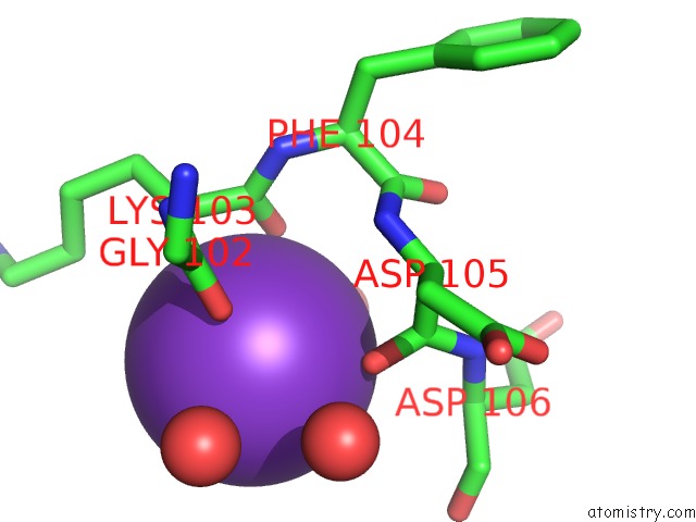
Mono view
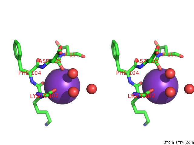
Stereo pair view
Mono view
Stereo pair view
A full contact list of Potassium with other atoms in the K binding
site number 1 of Crystal Structure of the 43K Atpase Domain of Escherichia Coli Gyrase B in Complex with An Aminocoumarin within 5.0Å range:
|
Potassium binding site 2 out of 3 in 6eng
Go back to
Potassium binding site 2 out
of 3 in the Crystal Structure of the 43K Atpase Domain of Escherichia Coli Gyrase B in Complex with An Aminocoumarin
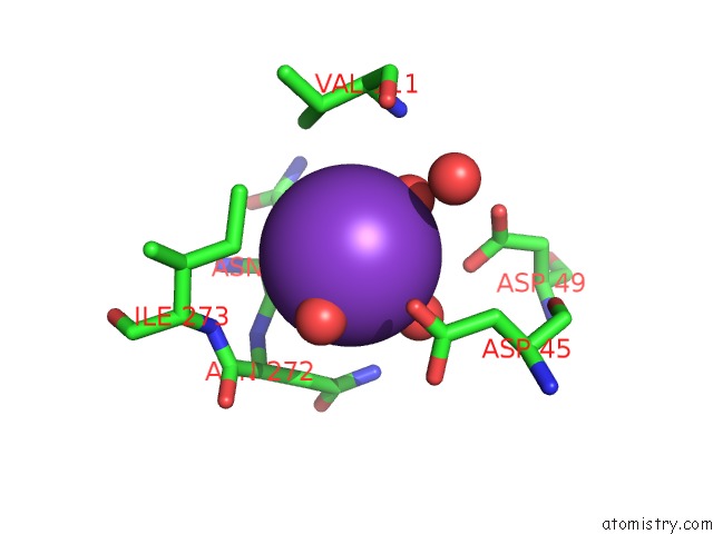
Mono view
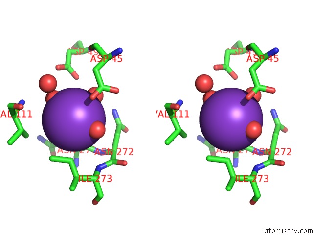
Stereo pair view
Mono view
Stereo pair view
A full contact list of Potassium with other atoms in the K binding
site number 2 of Crystal Structure of the 43K Atpase Domain of Escherichia Coli Gyrase B in Complex with An Aminocoumarin within 5.0Å range:
|
Potassium binding site 3 out of 3 in 6eng
Go back to
Potassium binding site 3 out
of 3 in the Crystal Structure of the 43K Atpase Domain of Escherichia Coli Gyrase B in Complex with An Aminocoumarin
Mono view
Stereo pair view
Mono view
Stereo pair view
A full contact list of Potassium with other atoms in the K binding
site number 3 of Crystal Structure of the 43K Atpase Domain of Escherichia Coli Gyrase B in Complex with An Aminocoumarin within 5.0Å range:
|
Reference:
A.Vanden Broeck,
A.G.Mcewen,
Y.Chebaro,
N.Potier,
V.Lamour.
Structural Basis For Dna Gyrase Interaction with Coumermycin A1. J.Med.Chem. V. 62 4225 2019.
ISSN: ISSN 0022-2623
PubMed: 30920824
DOI: 10.1021/ACS.JMEDCHEM.8B01928
Page generated: Sat Aug 9 10:53:48 2025
ISSN: ISSN 0022-2623
PubMed: 30920824
DOI: 10.1021/ACS.JMEDCHEM.8B01928
Last articles
K in 8CGIK in 8CG1
K in 8CFY
K in 8CG2
K in 8CFX
K in 8CG0
K in 8CFW
K in 8CFV
K in 8CFU
K in 8CFT