Potassium »
PDB 6dxz-6f3n »
6e8y »
Potassium in PDB 6e8y: Unliganded Human Glycerol 3-Phosphate Dehydrogenase
Enzymatic activity of Unliganded Human Glycerol 3-Phosphate Dehydrogenase
All present enzymatic activity of Unliganded Human Glycerol 3-Phosphate Dehydrogenase:
1.1.1.8;
1.1.1.8;
Protein crystallography data
The structure of Unliganded Human Glycerol 3-Phosphate Dehydrogenase, PDB code: 6e8y
was solved by
L.S.Mydy,
A.M.Gulick,
with X-Ray Crystallography technique. A brief refinement statistics is given in the table below:
| Resolution Low / High (Å) | 47.48 / 1.85 |
| Space group | P 1 21 1 |
| Cell size a, b, c (Å), α, β, γ (°) | 63.370, 76.160, 64.390, 90.00, 96.03, 90.00 |
| R / Rfree (%) | 17 / 21 |
Potassium Binding Sites:
The binding sites of Potassium atom in the Unliganded Human Glycerol 3-Phosphate Dehydrogenase
(pdb code 6e8y). This binding sites where shown within
5.0 Angstroms radius around Potassium atom.
In total 2 binding sites of Potassium where determined in the Unliganded Human Glycerol 3-Phosphate Dehydrogenase, PDB code: 6e8y:
Jump to Potassium binding site number: 1; 2;
In total 2 binding sites of Potassium where determined in the Unliganded Human Glycerol 3-Phosphate Dehydrogenase, PDB code: 6e8y:
Jump to Potassium binding site number: 1; 2;
Potassium binding site 1 out of 2 in 6e8y
Go back to
Potassium binding site 1 out
of 2 in the Unliganded Human Glycerol 3-Phosphate Dehydrogenase
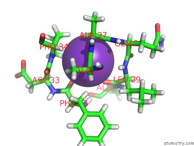
Mono view
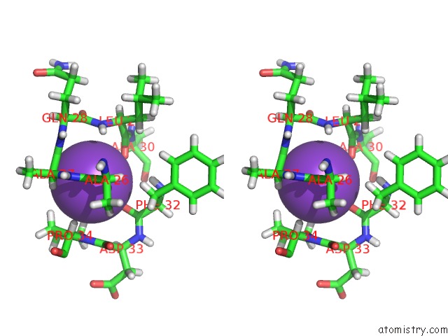
Stereo pair view
Mono view
Stereo pair view
A full contact list of Potassium with other atoms in the K binding
site number 1 of Unliganded Human Glycerol 3-Phosphate Dehydrogenase within 5.0Å range:
|
Potassium binding site 2 out of 2 in 6e8y
Go back to
Potassium binding site 2 out
of 2 in the Unliganded Human Glycerol 3-Phosphate Dehydrogenase
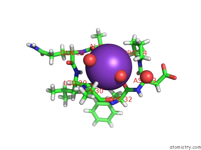
Mono view
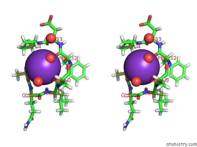
Stereo pair view
Mono view
Stereo pair view
A full contact list of Potassium with other atoms in the K binding
site number 2 of Unliganded Human Glycerol 3-Phosphate Dehydrogenase within 5.0Å range:
|
Reference:
L.S.Mydy,
J.R.Cristobal,
R.D.Katigbak,
P.Bauer,
A.C.Reyes,
S.C.L.Kamerlin,
J.P.Richard,
A.M.Gulick.
Human Glycerol 3-Phosphate Dehydrogenase: X-Ray Crystal Structures That Guide the Interpretation of Mutagenesis Studies. Biochemistry V. 58 1061 2019.
ISSN: ISSN 1520-4995
PubMed: 30640445
DOI: 10.1021/ACS.BIOCHEM.8B01103
Page generated: Mon Aug 12 15:58:56 2024
ISSN: ISSN 1520-4995
PubMed: 30640445
DOI: 10.1021/ACS.BIOCHEM.8B01103
Last articles
Zn in 9J0NZn in 9J0O
Zn in 9J0P
Zn in 9FJX
Zn in 9EKB
Zn in 9C0F
Zn in 9CAH
Zn in 9CH0
Zn in 9CH3
Zn in 9CH1