Potassium »
PDB 6doj-6dxv »
6dur »
Potassium in PDB 6dur: Citrobacter Freundii Tyrosine Phenol-Lyase Complexed with L- Phenylalanine
Enzymatic activity of Citrobacter Freundii Tyrosine Phenol-Lyase Complexed with L- Phenylalanine
All present enzymatic activity of Citrobacter Freundii Tyrosine Phenol-Lyase Complexed with L- Phenylalanine:
4.1.99.2;
4.1.99.2;
Protein crystallography data
The structure of Citrobacter Freundii Tyrosine Phenol-Lyase Complexed with L- Phenylalanine, PDB code: 6dur
was solved by
R.S.Phillips,
with X-Ray Crystallography technique. A brief refinement statistics is given in the table below:
| Resolution Low / High (Å) | 48.73 / 1.80 |
| Space group | P 2 21 21 |
| Cell size a, b, c (Å), α, β, γ (°) | 59.510, 133.330, 142.840, 90.00, 90.00, 90.00 |
| R / Rfree (%) | 17.2 / 19.3 |
Potassium Binding Sites:
The binding sites of Potassium atom in the Citrobacter Freundii Tyrosine Phenol-Lyase Complexed with L- Phenylalanine
(pdb code 6dur). This binding sites where shown within
5.0 Angstroms radius around Potassium atom.
In total 2 binding sites of Potassium where determined in the Citrobacter Freundii Tyrosine Phenol-Lyase Complexed with L- Phenylalanine, PDB code: 6dur:
Jump to Potassium binding site number: 1; 2;
In total 2 binding sites of Potassium where determined in the Citrobacter Freundii Tyrosine Phenol-Lyase Complexed with L- Phenylalanine, PDB code: 6dur:
Jump to Potassium binding site number: 1; 2;
Potassium binding site 1 out of 2 in 6dur
Go back to
Potassium binding site 1 out
of 2 in the Citrobacter Freundii Tyrosine Phenol-Lyase Complexed with L- Phenylalanine
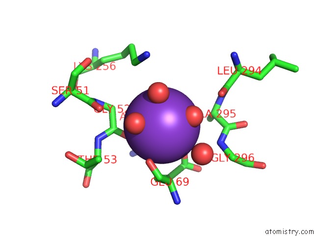
Mono view
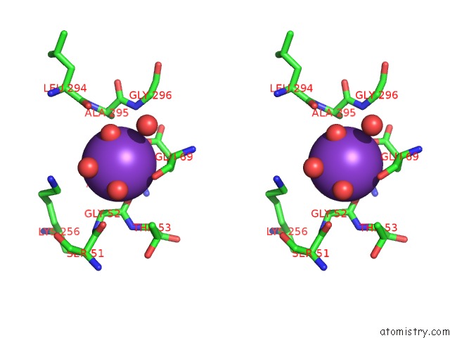
Stereo pair view
Mono view
Stereo pair view
A full contact list of Potassium with other atoms in the K binding
site number 1 of Citrobacter Freundii Tyrosine Phenol-Lyase Complexed with L- Phenylalanine within 5.0Å range:
|
Potassium binding site 2 out of 2 in 6dur
Go back to
Potassium binding site 2 out
of 2 in the Citrobacter Freundii Tyrosine Phenol-Lyase Complexed with L- Phenylalanine
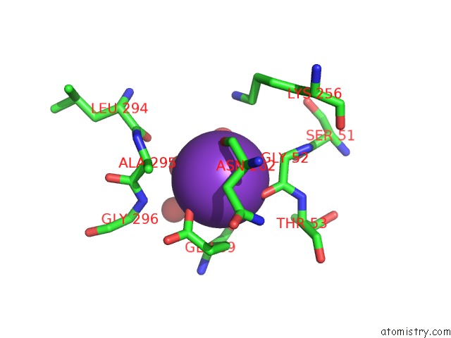
Mono view

Stereo pair view
Mono view
Stereo pair view
A full contact list of Potassium with other atoms in the K binding
site number 2 of Citrobacter Freundii Tyrosine Phenol-Lyase Complexed with L- Phenylalanine within 5.0Å range:
|
Reference:
R.S.Phillips,
S.Craig.
Crystal Structures of Wild-Type and F448A Mutant Citrobacter Freundii Tyrosine Phenol-Lyase Complexed with A Substrate and Inhibitors: Implications For the Reaction Mechanism. Biochemistry V. 57 6166 2018.
ISSN: ISSN 1520-4995
PubMed: 30260636
DOI: 10.1021/ACS.BIOCHEM.8B00724
Page generated: Mon Aug 12 15:54:26 2024
ISSN: ISSN 1520-4995
PubMed: 30260636
DOI: 10.1021/ACS.BIOCHEM.8B00724
Last articles
Zn in 9J0NZn in 9J0O
Zn in 9J0P
Zn in 9FJX
Zn in 9EKB
Zn in 9C0F
Zn in 9CAH
Zn in 9CH0
Zn in 9CH3
Zn in 9CH1