Potassium »
PDB 6css-6doi »
6doi »
Potassium in PDB 6doi: Crystal Structure of Bacillus Halodurans Ribonuclease H1 in Complex with An Rna/Dna Hybrid (1.54 Angstrom Wavelength): Soak in 0.5 Mm Egta and 200 Mm K+ at 21 C
Enzymatic activity of Crystal Structure of Bacillus Halodurans Ribonuclease H1 in Complex with An Rna/Dna Hybrid (1.54 Angstrom Wavelength): Soak in 0.5 Mm Egta and 200 Mm K+ at 21 C
All present enzymatic activity of Crystal Structure of Bacillus Halodurans Ribonuclease H1 in Complex with An Rna/Dna Hybrid (1.54 Angstrom Wavelength): Soak in 0.5 Mm Egta and 200 Mm K+ at 21 C:
3.1.26.4;
3.1.26.4;
Protein crystallography data
The structure of Crystal Structure of Bacillus Halodurans Ribonuclease H1 in Complex with An Rna/Dna Hybrid (1.54 Angstrom Wavelength): Soak in 0.5 Mm Egta and 200 Mm K+ at 21 C, PDB code: 6doi
was solved by
N.L.Samara,
W.Yang,
with X-Ray Crystallography technique. A brief refinement statistics is given in the table below:
| Resolution Low / High (Å) | 22.01 / 1.95 |
| Space group | C 1 2 1 |
| Cell size a, b, c (Å), α, β, γ (°) | 81.456, 38.020, 62.071, 90.00, 96.22, 90.00 |
| R / Rfree (%) | 14.7 / 19.5 |
Other elements in 6doi:
The structure of Crystal Structure of Bacillus Halodurans Ribonuclease H1 in Complex with An Rna/Dna Hybrid (1.54 Angstrom Wavelength): Soak in 0.5 Mm Egta and 200 Mm K+ at 21 C also contains other interesting chemical elements:
| Iodine | (I) | 4 atoms |
| Calcium | (Ca) | 1 atom |
Potassium Binding Sites:
The binding sites of Potassium atom in the Crystal Structure of Bacillus Halodurans Ribonuclease H1 in Complex with An Rna/Dna Hybrid (1.54 Angstrom Wavelength): Soak in 0.5 Mm Egta and 200 Mm K+ at 21 C
(pdb code 6doi). This binding sites where shown within
5.0 Angstroms radius around Potassium atom.
In total 3 binding sites of Potassium where determined in the Crystal Structure of Bacillus Halodurans Ribonuclease H1 in Complex with An Rna/Dna Hybrid (1.54 Angstrom Wavelength): Soak in 0.5 Mm Egta and 200 Mm K+ at 21 C, PDB code: 6doi:
Jump to Potassium binding site number: 1; 2; 3;
In total 3 binding sites of Potassium where determined in the Crystal Structure of Bacillus Halodurans Ribonuclease H1 in Complex with An Rna/Dna Hybrid (1.54 Angstrom Wavelength): Soak in 0.5 Mm Egta and 200 Mm K+ at 21 C, PDB code: 6doi:
Jump to Potassium binding site number: 1; 2; 3;
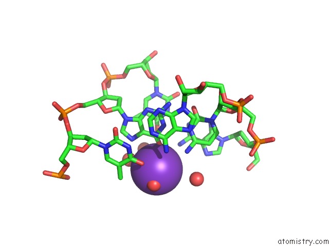
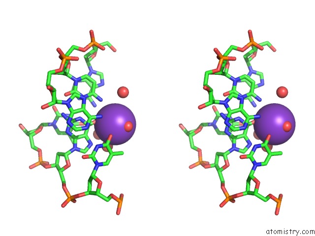
Potassium binding site 1 out of 3 in 6doi
Go back to
Potassium binding site 1 out
of 3 in the Crystal Structure of Bacillus Halodurans Ribonuclease H1 in Complex with An Rna/Dna Hybrid (1.54 Angstrom Wavelength): Soak in 0.5 Mm Egta and 200 Mm K+ at 21 C
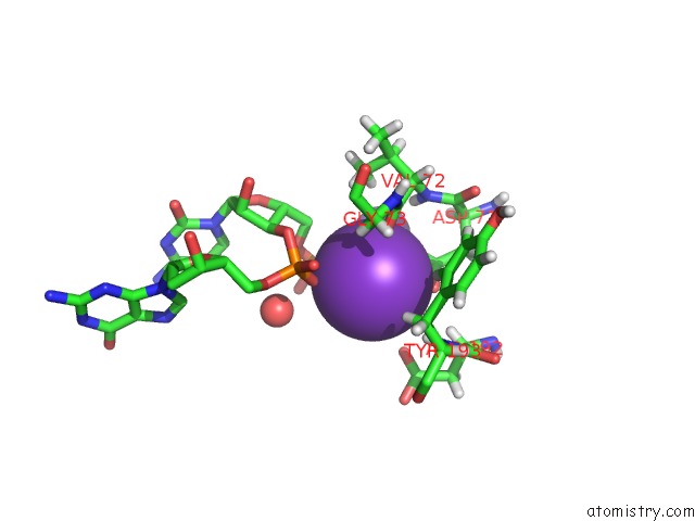
Mono view
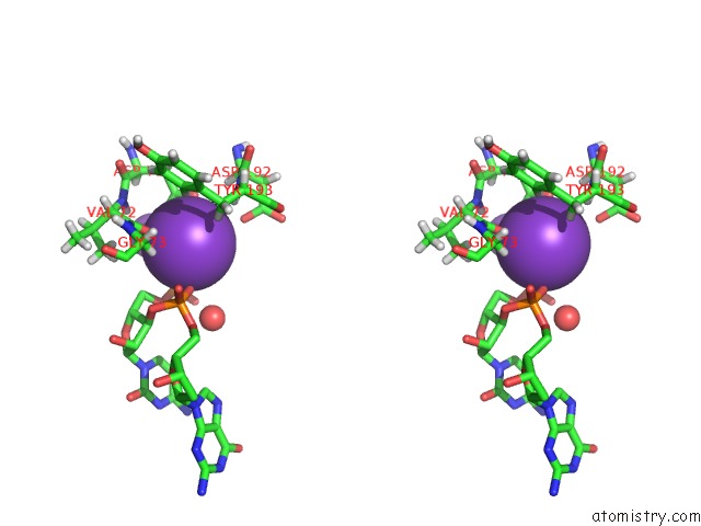
Stereo pair view
Mono view
Stereo pair view
A full contact list of Potassium with other atoms in the K binding
site number 1 of Crystal Structure of Bacillus Halodurans Ribonuclease H1 in Complex with An Rna/Dna Hybrid (1.54 Angstrom Wavelength): Soak in 0.5 Mm Egta and 200 Mm K+ at 21 C within 5.0Å range:
|
Potassium binding site 2 out of 3 in 6doi
Go back to
Potassium binding site 2 out
of 3 in the Crystal Structure of Bacillus Halodurans Ribonuclease H1 in Complex with An Rna/Dna Hybrid (1.54 Angstrom Wavelength): Soak in 0.5 Mm Egta and 200 Mm K+ at 21 C
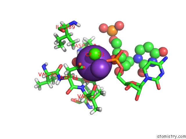
Mono view
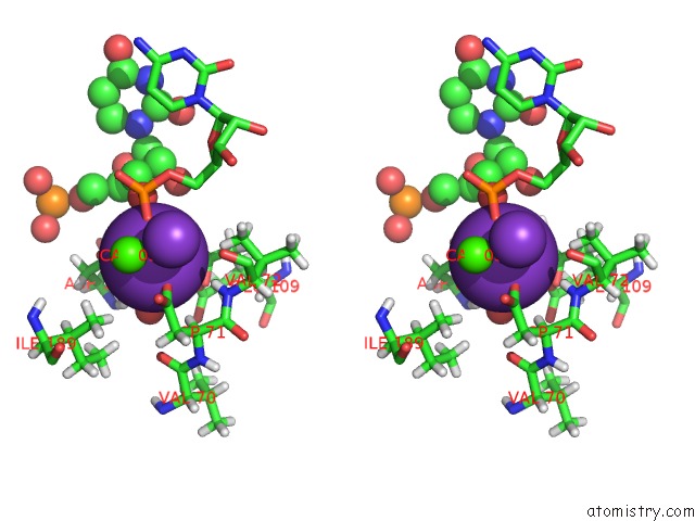
Stereo pair view
Mono view
Stereo pair view
A full contact list of Potassium with other atoms in the K binding
site number 2 of Crystal Structure of Bacillus Halodurans Ribonuclease H1 in Complex with An Rna/Dna Hybrid (1.54 Angstrom Wavelength): Soak in 0.5 Mm Egta and 200 Mm K+ at 21 C within 5.0Å range:
|
Potassium binding site 3 out of 3 in 6doi
Go back to
Potassium binding site 3 out
of 3 in the Crystal Structure of Bacillus Halodurans Ribonuclease H1 in Complex with An Rna/Dna Hybrid (1.54 Angstrom Wavelength): Soak in 0.5 Mm Egta and 200 Mm K+ at 21 C
Mono view
Stereo pair view
Mono view
Stereo pair view
A full contact list of Potassium with other atoms in the K binding
site number 3 of Crystal Structure of Bacillus Halodurans Ribonuclease H1 in Complex with An Rna/Dna Hybrid (1.54 Angstrom Wavelength): Soak in 0.5 Mm Egta and 200 Mm K+ at 21 C within 5.0Å range:
|
Reference:
N.L.Samara,
W.Yang.
Cation Trafficking Propels Rna Hydrolysis. Nat. Struct. Mol. Biol. V. 25 715 2018.
ISSN: ESSN 1545-9985
PubMed: 30076410
DOI: 10.1038/S41594-018-0099-4
Page generated: Mon Aug 12 15:48:39 2024
ISSN: ESSN 1545-9985
PubMed: 30076410
DOI: 10.1038/S41594-018-0099-4
Last articles
Zn in 9J0NZn in 9J0O
Zn in 9J0P
Zn in 9FJX
Zn in 9EKB
Zn in 9C0F
Zn in 9CAH
Zn in 9CH0
Zn in 9CH3
Zn in 9CH1