Potassium »
PDB 6css-6doi »
6css »
Potassium in PDB 6css: Crystal Structure of Danio Rerio Histone Deacetylase 6 Catalytic Domain 2 in Complex with Cyclopentenylhydroxamate
Protein crystallography data
The structure of Crystal Structure of Danio Rerio Histone Deacetylase 6 Catalytic Domain 2 in Complex with Cyclopentenylhydroxamate, PDB code: 6css
was solved by
N.J.Porter,
D.W.Christianson,
with X-Ray Crystallography technique. A brief refinement statistics is given in the table below:
| Resolution Low / High (Å) | 48.32 / 1.70 |
| Space group | P 21 21 21 |
| Cell size a, b, c (Å), α, β, γ (°) | 74.791, 92.021, 96.640, 90.00, 90.00, 90.00 |
| R / Rfree (%) | 17 / 19.3 |
Other elements in 6css:
The structure of Crystal Structure of Danio Rerio Histone Deacetylase 6 Catalytic Domain 2 in Complex with Cyclopentenylhydroxamate also contains other interesting chemical elements:
| Zinc | (Zn) | 2 atoms |
Potassium Binding Sites:
The binding sites of Potassium atom in the Crystal Structure of Danio Rerio Histone Deacetylase 6 Catalytic Domain 2 in Complex with Cyclopentenylhydroxamate
(pdb code 6css). This binding sites where shown within
5.0 Angstroms radius around Potassium atom.
In total 4 binding sites of Potassium where determined in the Crystal Structure of Danio Rerio Histone Deacetylase 6 Catalytic Domain 2 in Complex with Cyclopentenylhydroxamate, PDB code: 6css:
Jump to Potassium binding site number: 1; 2; 3; 4;
In total 4 binding sites of Potassium where determined in the Crystal Structure of Danio Rerio Histone Deacetylase 6 Catalytic Domain 2 in Complex with Cyclopentenylhydroxamate, PDB code: 6css:
Jump to Potassium binding site number: 1; 2; 3; 4;
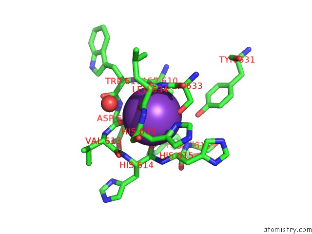
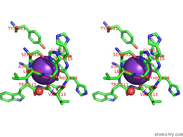
Potassium binding site 1 out of 4 in 6css
Go back to
Potassium binding site 1 out
of 4 in the Crystal Structure of Danio Rerio Histone Deacetylase 6 Catalytic Domain 2 in Complex with Cyclopentenylhydroxamate
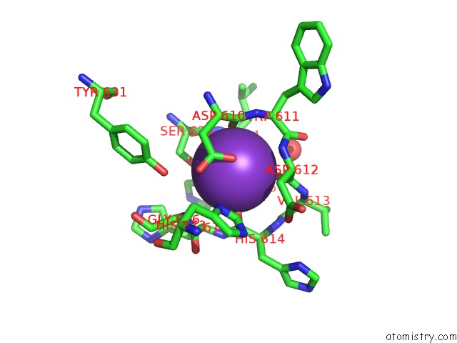
Mono view
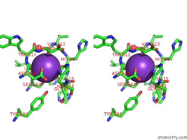
Stereo pair view
Mono view
Stereo pair view
A full contact list of Potassium with other atoms in the K binding
site number 1 of Crystal Structure of Danio Rerio Histone Deacetylase 6 Catalytic Domain 2 in Complex with Cyclopentenylhydroxamate within 5.0Å range:
|
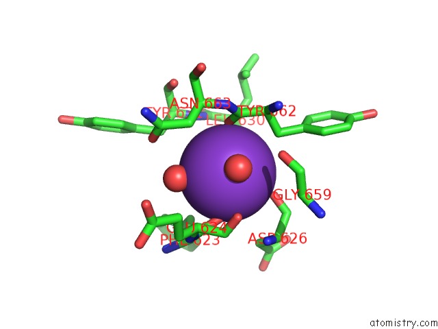
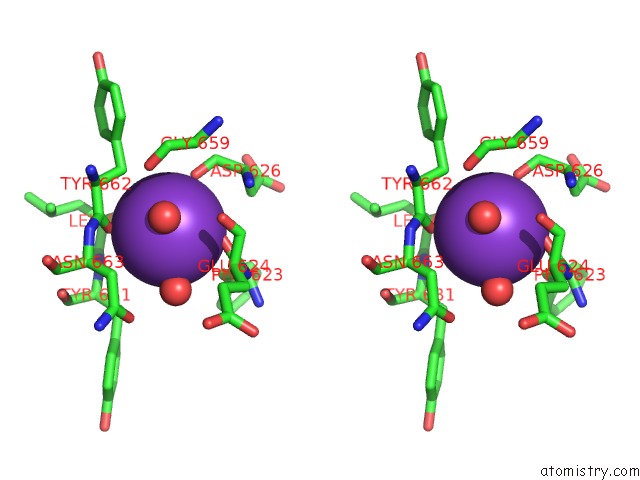
Potassium binding site 2 out of 4 in 6css
Go back to
Potassium binding site 2 out
of 4 in the Crystal Structure of Danio Rerio Histone Deacetylase 6 Catalytic Domain 2 in Complex with Cyclopentenylhydroxamate
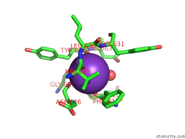
Mono view
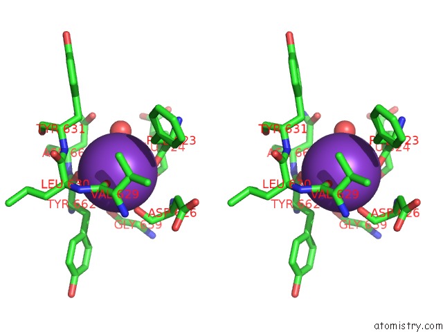
Stereo pair view
Mono view
Stereo pair view
A full contact list of Potassium with other atoms in the K binding
site number 2 of Crystal Structure of Danio Rerio Histone Deacetylase 6 Catalytic Domain 2 in Complex with Cyclopentenylhydroxamate within 5.0Å range:
|
Potassium binding site 3 out of 4 in 6css
Go back to
Potassium binding site 3 out
of 4 in the Crystal Structure of Danio Rerio Histone Deacetylase 6 Catalytic Domain 2 in Complex with Cyclopentenylhydroxamate
Mono view
Stereo pair view
Mono view
Stereo pair view
A full contact list of Potassium with other atoms in the K binding
site number 3 of Crystal Structure of Danio Rerio Histone Deacetylase 6 Catalytic Domain 2 in Complex with Cyclopentenylhydroxamate within 5.0Å range:
|
Potassium binding site 4 out of 4 in 6css
Go back to
Potassium binding site 4 out
of 4 in the Crystal Structure of Danio Rerio Histone Deacetylase 6 Catalytic Domain 2 in Complex with Cyclopentenylhydroxamate
Mono view
Stereo pair view
Mono view
Stereo pair view
A full contact list of Potassium with other atoms in the K binding
site number 4 of Crystal Structure of Danio Rerio Histone Deacetylase 6 Catalytic Domain 2 in Complex with Cyclopentenylhydroxamate within 5.0Å range:
|
Reference:
N.J.Porter,
F.F.Wagner,
D.W.Christianson.
Entropy As A Driver of Selectivity For Inhibitor Binding to Histone Deacetylase 6. Biochemistry V. 57 3916 2018.
ISSN: ISSN 1520-4995
PubMed: 29775292
DOI: 10.1021/ACS.BIOCHEM.8B00367
Page generated: Mon Aug 12 15:42:36 2024
ISSN: ISSN 1520-4995
PubMed: 29775292
DOI: 10.1021/ACS.BIOCHEM.8B00367
Last articles
Zn in 9MJ5Zn in 9HNW
Zn in 9G0L
Zn in 9FNE
Zn in 9DZN
Zn in 9E0I
Zn in 9D32
Zn in 9DAK
Zn in 8ZXC
Zn in 8ZUF