Potassium »
PDB 5zod-6b7f »
6b7d »
Potassium in PDB 6b7d: Crystal Structure of E.Coli Phosphopantetheine Adenylyltransferase (Ppat/Coad) in Complex with 3-(4-Chlorophenyl)-6-Methoxy-4,5- Dimethylpyridazine
Enzymatic activity of Crystal Structure of E.Coli Phosphopantetheine Adenylyltransferase (Ppat/Coad) in Complex with 3-(4-Chlorophenyl)-6-Methoxy-4,5- Dimethylpyridazine
All present enzymatic activity of Crystal Structure of E.Coli Phosphopantetheine Adenylyltransferase (Ppat/Coad) in Complex with 3-(4-Chlorophenyl)-6-Methoxy-4,5- Dimethylpyridazine:
2.7.7.3;
2.7.7.3;
Protein crystallography data
The structure of Crystal Structure of E.Coli Phosphopantetheine Adenylyltransferase (Ppat/Coad) in Complex with 3-(4-Chlorophenyl)-6-Methoxy-4,5- Dimethylpyridazine, PDB code: 6b7d
was solved by
A.W.Proudfoot,
D.Bussiere,
A.Lingel,
with X-Ray Crystallography technique. A brief refinement statistics is given in the table below:
| Resolution Low / High (Å) | 28.78 / 1.80 |
| Space group | I 2 3 |
| Cell size a, b, c (Å), α, β, γ (°) | 134.990, 134.990, 134.990, 90.00, 90.00, 90.00 |
| R / Rfree (%) | 18.4 / 19.7 |
Other elements in 6b7d:
The structure of Crystal Structure of E.Coli Phosphopantetheine Adenylyltransferase (Ppat/Coad) in Complex with 3-(4-Chlorophenyl)-6-Methoxy-4,5- Dimethylpyridazine also contains other interesting chemical elements:
| Chlorine | (Cl) | 2 atoms |
Potassium Binding Sites:
The binding sites of Potassium atom in the Crystal Structure of E.Coli Phosphopantetheine Adenylyltransferase (Ppat/Coad) in Complex with 3-(4-Chlorophenyl)-6-Methoxy-4,5- Dimethylpyridazine
(pdb code 6b7d). This binding sites where shown within
5.0 Angstroms radius around Potassium atom.
In total 2 binding sites of Potassium where determined in the Crystal Structure of E.Coli Phosphopantetheine Adenylyltransferase (Ppat/Coad) in Complex with 3-(4-Chlorophenyl)-6-Methoxy-4,5- Dimethylpyridazine, PDB code: 6b7d:
Jump to Potassium binding site number: 1; 2;
In total 2 binding sites of Potassium where determined in the Crystal Structure of E.Coli Phosphopantetheine Adenylyltransferase (Ppat/Coad) in Complex with 3-(4-Chlorophenyl)-6-Methoxy-4,5- Dimethylpyridazine, PDB code: 6b7d:
Jump to Potassium binding site number: 1; 2;
Potassium binding site 1 out of 2 in 6b7d
Go back to
Potassium binding site 1 out
of 2 in the Crystal Structure of E.Coli Phosphopantetheine Adenylyltransferase (Ppat/Coad) in Complex with 3-(4-Chlorophenyl)-6-Methoxy-4,5- Dimethylpyridazine
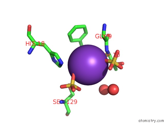
Mono view
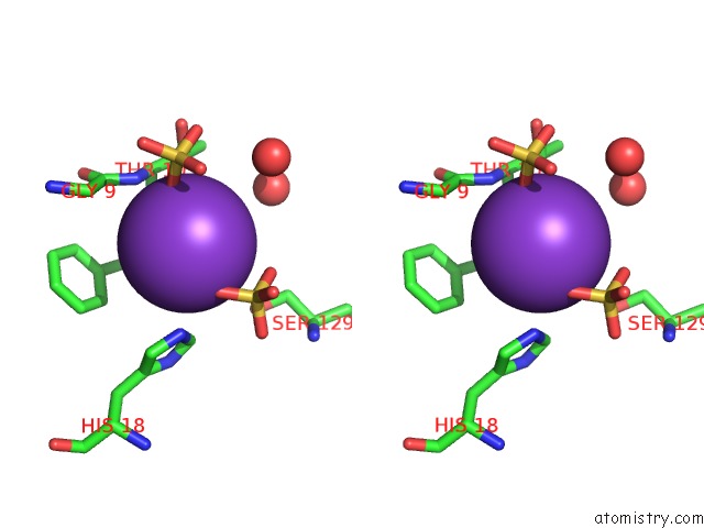
Stereo pair view
Mono view
Stereo pair view
A full contact list of Potassium with other atoms in the K binding
site number 1 of Crystal Structure of E.Coli Phosphopantetheine Adenylyltransferase (Ppat/Coad) in Complex with 3-(4-Chlorophenyl)-6-Methoxy-4,5- Dimethylpyridazine within 5.0Å range:
|
Potassium binding site 2 out of 2 in 6b7d
Go back to
Potassium binding site 2 out
of 2 in the Crystal Structure of E.Coli Phosphopantetheine Adenylyltransferase (Ppat/Coad) in Complex with 3-(4-Chlorophenyl)-6-Methoxy-4,5- Dimethylpyridazine
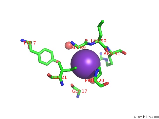
Mono view
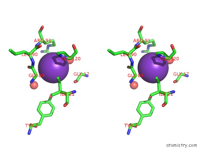
Stereo pair view
Mono view
Stereo pair view
A full contact list of Potassium with other atoms in the K binding
site number 2 of Crystal Structure of E.Coli Phosphopantetheine Adenylyltransferase (Ppat/Coad) in Complex with 3-(4-Chlorophenyl)-6-Methoxy-4,5- Dimethylpyridazine within 5.0Å range:
|
Reference:
A.Proudfoot,
D.E.Bussiere,
A.Lingel.
High-Confidence Protein-Ligand Complex Modeling By uc(Nmr)-Guided Docking Enables Early Hit Optimization. J. Am. Chem. Soc. V. 139 17824 2017.
ISSN: ESSN 1520-5126
PubMed: 29190085
DOI: 10.1021/JACS.7B07171
Page generated: Mon Aug 12 15:25:52 2024
ISSN: ESSN 1520-5126
PubMed: 29190085
DOI: 10.1021/JACS.7B07171
Last articles
Zn in 9JYWZn in 9IR4
Zn in 9IR3
Zn in 9GMX
Zn in 9GMW
Zn in 9JEJ
Zn in 9ERF
Zn in 9ERE
Zn in 9EGV
Zn in 9EGW