Potassium »
PDB 5zod-6b7f »
6aao »
Potassium in PDB 6aao: Crystal Structure of Methanosarcina Mazei Pylrs(Y306A/Y384F) Complexed with Tco*Lys
Enzymatic activity of Crystal Structure of Methanosarcina Mazei Pylrs(Y306A/Y384F) Complexed with Tco*Lys
All present enzymatic activity of Crystal Structure of Methanosarcina Mazei Pylrs(Y306A/Y384F) Complexed with Tco*Lys:
6.1.1.26;
6.1.1.26;
Protein crystallography data
The structure of Crystal Structure of Methanosarcina Mazei Pylrs(Y306A/Y384F) Complexed with Tco*Lys, PDB code: 6aao
was solved by
T.Yanagisawa,
M.Kuratani,
S.Yokoyama,
with X-Ray Crystallography technique. A brief refinement statistics is given in the table below:
| Resolution Low / High (Å) | 35.13 / 1.40 |
| Space group | C 1 2 1 |
| Cell size a, b, c (Å), α, β, γ (°) | 101.588, 43.971, 72.739, 90.00, 118.72, 90.00 |
| R / Rfree (%) | 15.2 / 19.6 |
Other elements in 6aao:
The structure of Crystal Structure of Methanosarcina Mazei Pylrs(Y306A/Y384F) Complexed with Tco*Lys also contains other interesting chemical elements:
| Magnesium | (Mg) | 3 atoms |
Potassium Binding Sites:
The binding sites of Potassium atom in the Crystal Structure of Methanosarcina Mazei Pylrs(Y306A/Y384F) Complexed with Tco*Lys
(pdb code 6aao). This binding sites where shown within
5.0 Angstroms radius around Potassium atom.
In total 2 binding sites of Potassium where determined in the Crystal Structure of Methanosarcina Mazei Pylrs(Y306A/Y384F) Complexed with Tco*Lys, PDB code: 6aao:
Jump to Potassium binding site number: 1; 2;
In total 2 binding sites of Potassium where determined in the Crystal Structure of Methanosarcina Mazei Pylrs(Y306A/Y384F) Complexed with Tco*Lys, PDB code: 6aao:
Jump to Potassium binding site number: 1; 2;
Potassium binding site 1 out of 2 in 6aao
Go back to
Potassium binding site 1 out
of 2 in the Crystal Structure of Methanosarcina Mazei Pylrs(Y306A/Y384F) Complexed with Tco*Lys
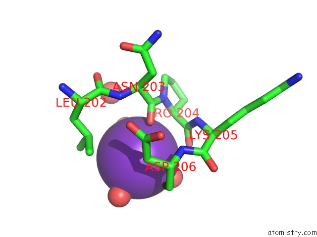
Mono view
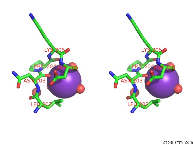
Stereo pair view
Mono view
Stereo pair view
A full contact list of Potassium with other atoms in the K binding
site number 1 of Crystal Structure of Methanosarcina Mazei Pylrs(Y306A/Y384F) Complexed with Tco*Lys within 5.0Å range:
|
Potassium binding site 2 out of 2 in 6aao
Go back to
Potassium binding site 2 out
of 2 in the Crystal Structure of Methanosarcina Mazei Pylrs(Y306A/Y384F) Complexed with Tco*Lys
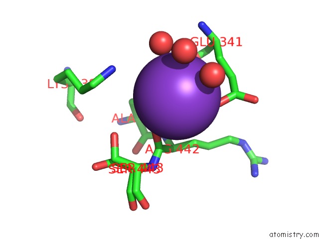
Mono view
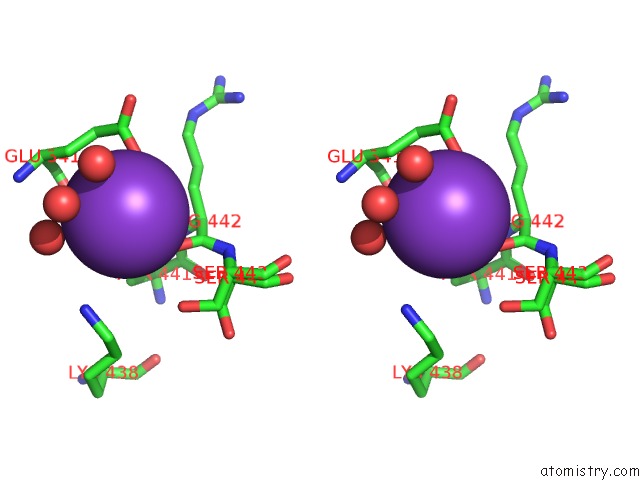
Stereo pair view
Mono view
Stereo pair view
A full contact list of Potassium with other atoms in the K binding
site number 2 of Crystal Structure of Methanosarcina Mazei Pylrs(Y306A/Y384F) Complexed with Tco*Lys within 5.0Å range:
|
Reference:
T.Yanagisawa,
M.Kuratani,
E.Seki,
N.Hino,
K.Sakamoto,
S.Yokoyama.
Structural Basis For Genetic-Code Expansion with Bulky Lysine Derivatives By An Engineered Pyrrolysyl-Trna Synthetase. Cell Chem Biol V. 26 936 2019.
ISSN: ESSN 2451-9456
PubMed: 31031143
DOI: 10.1016/J.CHEMBIOL.2019.03.008
Page generated: Mon Aug 12 15:19:12 2024
ISSN: ESSN 2451-9456
PubMed: 31031143
DOI: 10.1016/J.CHEMBIOL.2019.03.008
Last articles
Zn in 9J0NZn in 9J0O
Zn in 9J0P
Zn in 9FJX
Zn in 9EKB
Zn in 9C0F
Zn in 9CAH
Zn in 9CH0
Zn in 9CH3
Zn in 9CH1