Potassium »
PDB 5x24-5ze2 »
5yew »
Potassium in PDB 5yew: Structural Basis For Gtp Hydrolysis and Conformational Change of Mitofusin 1 in Mediating Mitochondrial Fusion
Protein crystallography data
The structure of Structural Basis For Gtp Hydrolysis and Conformational Change of Mitofusin 1 in Mediating Mitochondrial Fusion, PDB code: 5yew
was solved by
L.Yan,
Y.Qi,
X.Huang,
C.Yu,
with X-Ray Crystallography technique. A brief refinement statistics is given in the table below:
| Resolution Low / High (Å) | 50.00 / 3.20 |
| Space group | P 32 2 1 |
| Cell size a, b, c (Å), α, β, γ (°) | 207.942, 207.942, 107.893, 90.00, 90.00, 120.00 |
| R / Rfree (%) | 22.3 / 25.7 |
Other elements in 5yew:
The structure of Structural Basis For Gtp Hydrolysis and Conformational Change of Mitofusin 1 in Mediating Mitochondrial Fusion also contains other interesting chemical elements:
| Fluorine | (F) | 9 atoms |
| Magnesium | (Mg) | 3 atoms |
Potassium Binding Sites:
The binding sites of Potassium atom in the Structural Basis For Gtp Hydrolysis and Conformational Change of Mitofusin 1 in Mediating Mitochondrial Fusion
(pdb code 5yew). This binding sites where shown within
5.0 Angstroms radius around Potassium atom.
In total 3 binding sites of Potassium where determined in the Structural Basis For Gtp Hydrolysis and Conformational Change of Mitofusin 1 in Mediating Mitochondrial Fusion, PDB code: 5yew:
Jump to Potassium binding site number: 1; 2; 3;
In total 3 binding sites of Potassium where determined in the Structural Basis For Gtp Hydrolysis and Conformational Change of Mitofusin 1 in Mediating Mitochondrial Fusion, PDB code: 5yew:
Jump to Potassium binding site number: 1; 2; 3;
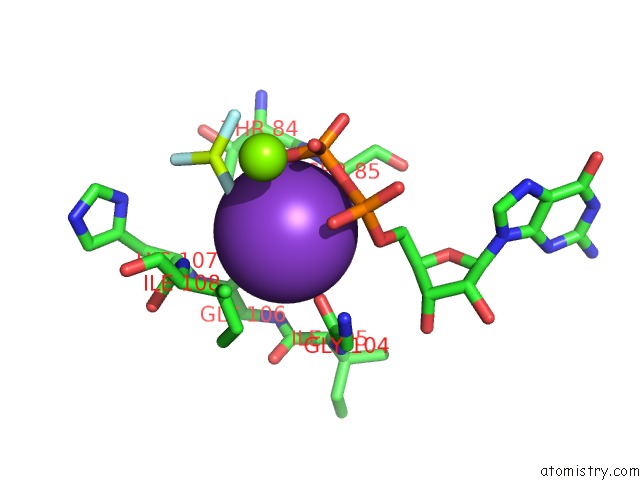
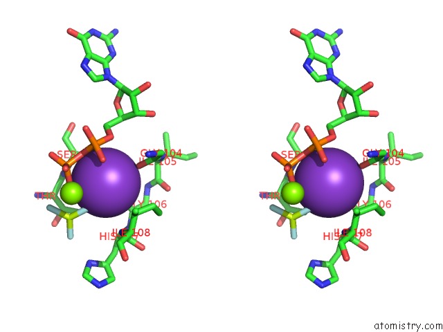
Potassium binding site 1 out of 3 in 5yew
Go back to
Potassium binding site 1 out
of 3 in the Structural Basis For Gtp Hydrolysis and Conformational Change of Mitofusin 1 in Mediating Mitochondrial Fusion
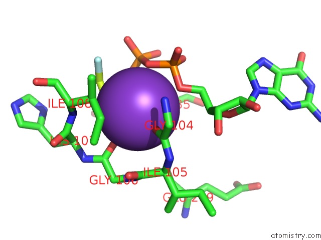
Mono view
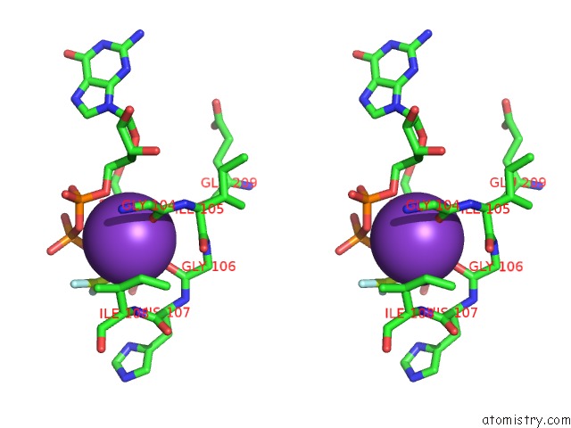
Stereo pair view
Mono view
Stereo pair view
A full contact list of Potassium with other atoms in the K binding
site number 1 of Structural Basis For Gtp Hydrolysis and Conformational Change of Mitofusin 1 in Mediating Mitochondrial Fusion within 5.0Å range:
|
Potassium binding site 2 out of 3 in 5yew
Go back to
Potassium binding site 2 out
of 3 in the Structural Basis For Gtp Hydrolysis and Conformational Change of Mitofusin 1 in Mediating Mitochondrial Fusion
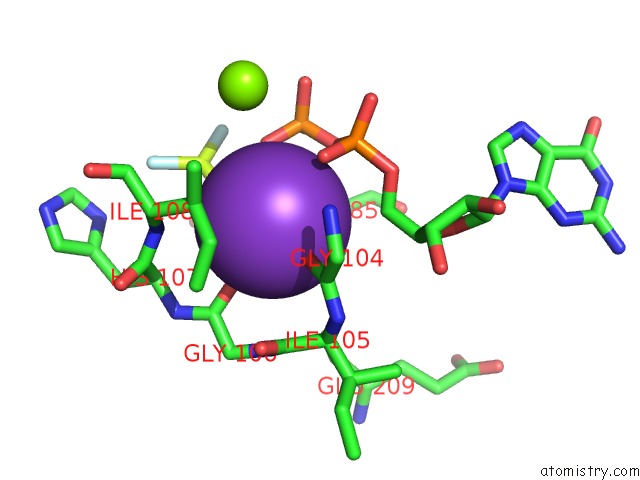
Mono view
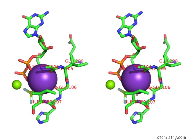
Stereo pair view
Mono view
Stereo pair view
A full contact list of Potassium with other atoms in the K binding
site number 2 of Structural Basis For Gtp Hydrolysis and Conformational Change of Mitofusin 1 in Mediating Mitochondrial Fusion within 5.0Å range:
|
Potassium binding site 3 out of 3 in 5yew
Go back to
Potassium binding site 3 out
of 3 in the Structural Basis For Gtp Hydrolysis and Conformational Change of Mitofusin 1 in Mediating Mitochondrial Fusion
Mono view
Stereo pair view
Mono view
Stereo pair view
A full contact list of Potassium with other atoms in the K binding
site number 3 of Structural Basis For Gtp Hydrolysis and Conformational Change of Mitofusin 1 in Mediating Mitochondrial Fusion within 5.0Å range:
|
Reference:
L.Yan,
Y.Qi,
X.Huang,
C.Yu,
L.Lan,
X.Guo,
Z.Rao,
J.Hu,
Z.Lou.
Structural Basis For Gtp Hydrolysis and Conformational Change of MFN1 in Mediating Membrane Fusion Nat. Struct. Mol. Biol. V. 25 233 2018.
ISSN: ESSN 1545-9985
PubMed: 29483649
DOI: 10.1038/S41594-018-0034-8
Page generated: Mon Aug 12 15:10:14 2024
ISSN: ESSN 1545-9985
PubMed: 29483649
DOI: 10.1038/S41594-018-0034-8
Last articles
Zn in 9J0NZn in 9J0O
Zn in 9J0P
Zn in 9FJX
Zn in 9EKB
Zn in 9C0F
Zn in 9CAH
Zn in 9CH0
Zn in 9CH3
Zn in 9CH1