Potassium »
PDB 5x24-5ze2 »
5xj7 »
Potassium in PDB 5xj7: Crystal Structure of Plsy (Ygih), An Integral Membrane Glycerol 3- Phosphate Acyltransferase - the Acyl Phosphate Form
Protein crystallography data
The structure of Crystal Structure of Plsy (Ygih), An Integral Membrane Glycerol 3- Phosphate Acyltransferase - the Acyl Phosphate Form, PDB code: 5xj7
was solved by
Y.Tang,
Z.Li,
D.Li,
with X-Ray Crystallography technique. A brief refinement statistics is given in the table below:
| Resolution Low / High (Å) | 40.57 / 1.77 |
| Space group | P 21 21 21 |
| Cell size a, b, c (Å), α, β, γ (°) | 46.224, 65.599, 84.678, 90.00, 90.00, 90.00 |
| R / Rfree (%) | 19.5 / 22.5 |
Potassium Binding Sites:
The binding sites of Potassium atom in the Crystal Structure of Plsy (Ygih), An Integral Membrane Glycerol 3- Phosphate Acyltransferase - the Acyl Phosphate Form
(pdb code 5xj7). This binding sites where shown within
5.0 Angstroms radius around Potassium atom.
In total 2 binding sites of Potassium where determined in the Crystal Structure of Plsy (Ygih), An Integral Membrane Glycerol 3- Phosphate Acyltransferase - the Acyl Phosphate Form, PDB code: 5xj7:
Jump to Potassium binding site number: 1; 2;
In total 2 binding sites of Potassium where determined in the Crystal Structure of Plsy (Ygih), An Integral Membrane Glycerol 3- Phosphate Acyltransferase - the Acyl Phosphate Form, PDB code: 5xj7:
Jump to Potassium binding site number: 1; 2;
Potassium binding site 1 out of 2 in 5xj7
Go back to
Potassium binding site 1 out
of 2 in the Crystal Structure of Plsy (Ygih), An Integral Membrane Glycerol 3- Phosphate Acyltransferase - the Acyl Phosphate Form
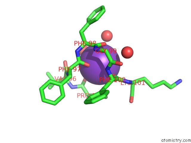
Mono view
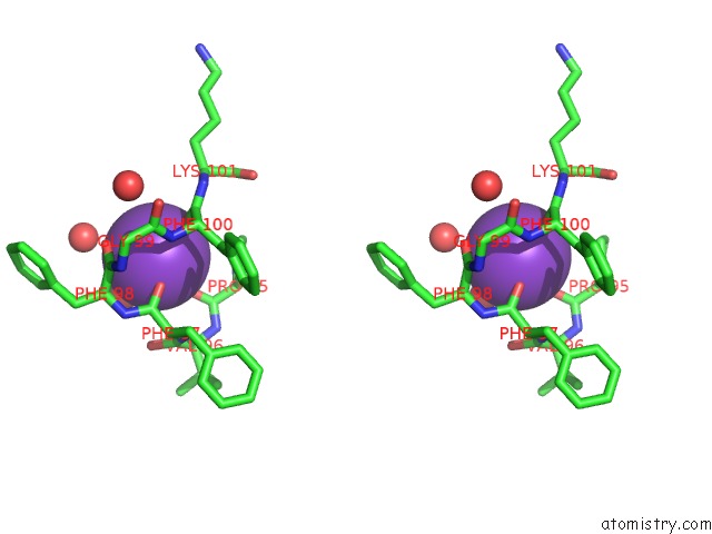
Stereo pair view
Mono view
Stereo pair view
A full contact list of Potassium with other atoms in the K binding
site number 1 of Crystal Structure of Plsy (Ygih), An Integral Membrane Glycerol 3- Phosphate Acyltransferase - the Acyl Phosphate Form within 5.0Å range:
|
Potassium binding site 2 out of 2 in 5xj7
Go back to
Potassium binding site 2 out
of 2 in the Crystal Structure of Plsy (Ygih), An Integral Membrane Glycerol 3- Phosphate Acyltransferase - the Acyl Phosphate Form
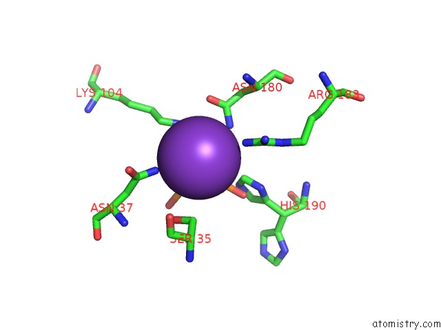
Mono view
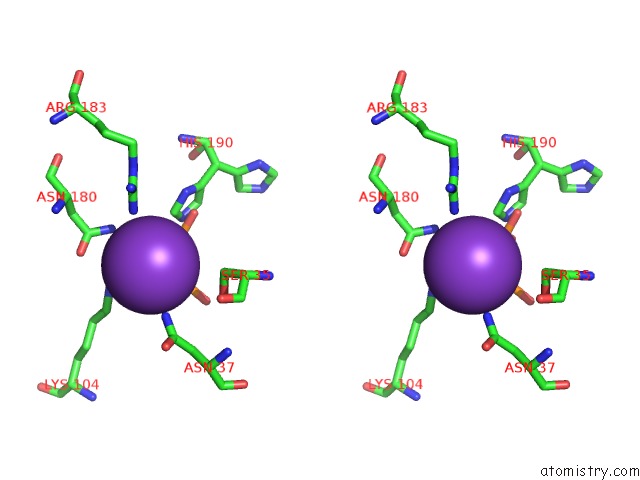
Stereo pair view
Mono view
Stereo pair view
A full contact list of Potassium with other atoms in the K binding
site number 2 of Crystal Structure of Plsy (Ygih), An Integral Membrane Glycerol 3- Phosphate Acyltransferase - the Acyl Phosphate Form within 5.0Å range:
|
Reference:
Z.Li,
Y.Tang,
Y.Wu,
S.Zhao,
J.Bao,
Y.Luo,
D.Li.
Structural Insights Into the Committed Step of Bacterial Phospholipid Biosynthesis. Nat Commun V. 8 1691 2017.
ISSN: ESSN 2041-1723
PubMed: 29167463
DOI: 10.1038/S41467-017-01821-9
Page generated: Mon Aug 12 15:09:24 2024
ISSN: ESSN 2041-1723
PubMed: 29167463
DOI: 10.1038/S41467-017-01821-9
Last articles
Zn in 9MJ5Zn in 9HNW
Zn in 9G0L
Zn in 9FNE
Zn in 9DZN
Zn in 9E0I
Zn in 9D32
Zn in 9DAK
Zn in 8ZXC
Zn in 8ZUF