Potassium »
PDB 5vt3-5x23 »
5x23 »
Potassium in PDB 5x23: Crystal Structure of CYP2C9 Genetic Variant A477T (*30) in Complex with Multiple Losartan Molecules
Enzymatic activity of Crystal Structure of CYP2C9 Genetic Variant A477T (*30) in Complex with Multiple Losartan Molecules
All present enzymatic activity of Crystal Structure of CYP2C9 Genetic Variant A477T (*30) in Complex with Multiple Losartan Molecules:
1.14.13.48; 1.14.13.49; 1.14.13.80; 1.14.99.38;
1.14.13.48; 1.14.13.49; 1.14.13.80; 1.14.99.38;
Protein crystallography data
The structure of Crystal Structure of CYP2C9 Genetic Variant A477T (*30) in Complex with Multiple Losartan Molecules, PDB code: 5x23
was solved by
K.Maekawa,
M.Adachi,
M.B.Shah,
with X-Ray Crystallography technique. A brief refinement statistics is given in the table below:
| Resolution Low / High (Å) | 45.51 / 2.00 |
| Space group | I 2 2 2 |
| Cell size a, b, c (Å), α, β, γ (°) | 74.952, 142.273, 161.831, 90.00, 90.00, 90.00 |
| R / Rfree (%) | 21.7 / 25.3 |
Other elements in 5x23:
The structure of Crystal Structure of CYP2C9 Genetic Variant A477T (*30) in Complex with Multiple Losartan Molecules also contains other interesting chemical elements:
| Iron | (Fe) | 1 atom |
| Chlorine | (Cl) | 4 atoms |
Potassium Binding Sites:
The binding sites of Potassium atom in the Crystal Structure of CYP2C9 Genetic Variant A477T (*30) in Complex with Multiple Losartan Molecules
(pdb code 5x23). This binding sites where shown within
5.0 Angstroms radius around Potassium atom.
In total only one binding site of Potassium was determined in the Crystal Structure of CYP2C9 Genetic Variant A477T (*30) in Complex with Multiple Losartan Molecules, PDB code: 5x23:
In total only one binding site of Potassium was determined in the Crystal Structure of CYP2C9 Genetic Variant A477T (*30) in Complex with Multiple Losartan Molecules, PDB code: 5x23:
Potassium binding site 1 out of 1 in 5x23
Go back to
Potassium binding site 1 out
of 1 in the Crystal Structure of CYP2C9 Genetic Variant A477T (*30) in Complex with Multiple Losartan Molecules
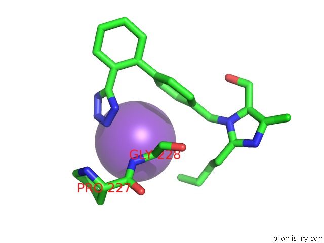
Mono view
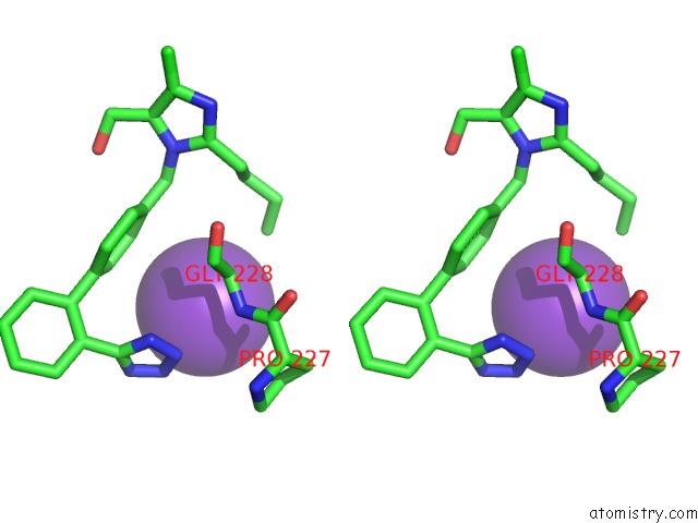
Stereo pair view
Mono view
Stereo pair view
A full contact list of Potassium with other atoms in the K binding
site number 1 of Crystal Structure of CYP2C9 Genetic Variant A477T (*30) in Complex with Multiple Losartan Molecules within 5.0Å range:
|
Reference:
K.Maekawa,
M.Adachi,
Y.Matsuzawa,
Q.Zhang,
R.Kuroki,
Y.Saito,
M.B.Shah.
Structural Basis of Single-Nucleotide Polymorphisms in Cytochrome P450 2C9 Biochemistry V. 56 5476 2017.
ISSN: ISSN 1520-4995
PubMed: 28972767
DOI: 10.1021/ACS.BIOCHEM.7B00795
Page generated: Sat Aug 9 10:11:54 2025
ISSN: ISSN 1520-4995
PubMed: 28972767
DOI: 10.1021/ACS.BIOCHEM.7B00795
Last articles
Mg in 2HO4Mg in 2HNY
Mg in 2HMF
Mg in 2HMC
Mg in 2HND
Mg in 2HMA
Mg in 2HJ6
Mg in 2HK6
Mg in 2HKJ
Mg in 2HIT