Potassium »
PDB 5vt3-5x23 »
5wjm »
Potassium in PDB 5wjm: Crystal Structure of Mouse Cadherin-23 EC17-18
Protein crystallography data
The structure of Crystal Structure of Mouse Cadherin-23 EC17-18, PDB code: 5wjm
was solved by
P.De-La-Torre,
M.Sotomayor,
with X-Ray Crystallography technique. A brief refinement statistics is given in the table below:
| Resolution Low / High (Å) | 86.60 / 2.90 |
| Space group | P 43 21 2 |
| Cell size a, b, c (Å), α, β, γ (°) | 122.476, 122.476, 43.603, 90.00, 90.00, 90.00 |
| R / Rfree (%) | 20 / 26.6 |
Other elements in 5wjm:
The structure of Crystal Structure of Mouse Cadherin-23 EC17-18 also contains other interesting chemical elements:
| Calcium | (Ca) | 6 atoms |
Potassium Binding Sites:
The binding sites of Potassium atom in the Crystal Structure of Mouse Cadherin-23 EC17-18
(pdb code 5wjm). This binding sites where shown within
5.0 Angstroms radius around Potassium atom.
In total only one binding site of Potassium was determined in the Crystal Structure of Mouse Cadherin-23 EC17-18, PDB code: 5wjm:
In total only one binding site of Potassium was determined in the Crystal Structure of Mouse Cadherin-23 EC17-18, PDB code: 5wjm:
Potassium binding site 1 out of 1 in 5wjm
Go back to
Potassium binding site 1 out
of 1 in the Crystal Structure of Mouse Cadherin-23 EC17-18
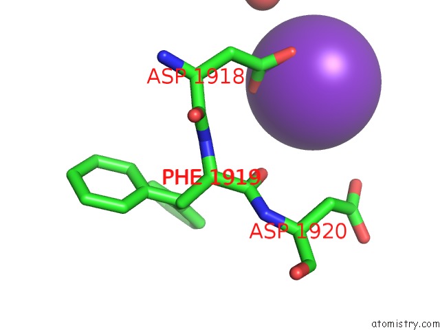
Mono view
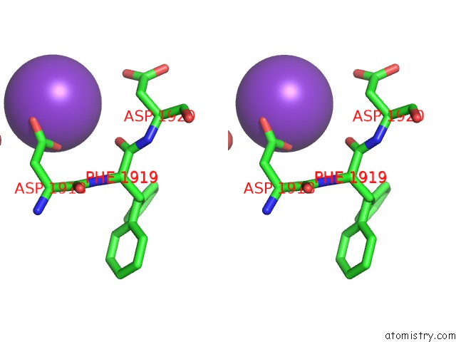
Stereo pair view
Mono view
Stereo pair view
A full contact list of Potassium with other atoms in the K binding
site number 1 of Crystal Structure of Mouse Cadherin-23 EC17-18 within 5.0Å range:
|
Reference:
A.Jaiganesh,
P.De-La-Torre,
A.A.Patel,
D.J.Termine,
F.Velez-Cortes,
C.Chen,
M.Sotomayor.
Zooming in on Cadherin-23: Structural Diversity and Potential Mechanisms of Inherited Deafness. Structure V. 26 1210 2018.
ISSN: ISSN 1878-4186
PubMed: 30033219
DOI: 10.1016/J.STR.2018.06.003
Page generated: Sat Aug 9 10:06:05 2025
ISSN: ISSN 1878-4186
PubMed: 30033219
DOI: 10.1016/J.STR.2018.06.003
Last articles
Mg in 1YLSMg in 1YL7
Mg in 1YL6
Mg in 1YL5
Mg in 1YKV
Mg in 1YKQ
Mg in 1YIT
Mg in 1YIJ
Mg in 1YJF
Mg in 1YI2