Potassium »
PDB 5vt3-5x23 »
5wgm »
Potassium in PDB 5wgm: Crystal Structure of Danio Rerio Histone Deacetylase 6 Catalytic Domain 2 in Complex with Acy-1083
Protein crystallography data
The structure of Crystal Structure of Danio Rerio Histone Deacetylase 6 Catalytic Domain 2 in Complex with Acy-1083, PDB code: 5wgm
was solved by
N.J.Porter,
D.W.Christianson,
with X-Ray Crystallography technique. A brief refinement statistics is given in the table below:
| Resolution Low / High (Å) | 49.95 / 1.75 |
| Space group | C 2 2 21 |
| Cell size a, b, c (Å), α, β, γ (°) | 97.813, 174.304, 149.064, 90.00, 90.00, 90.00 |
| R / Rfree (%) | 15.9 / 18.5 |
Other elements in 5wgm:
The structure of Crystal Structure of Danio Rerio Histone Deacetylase 6 Catalytic Domain 2 in Complex with Acy-1083 also contains other interesting chemical elements:
| Fluorine | (F) | 6 atoms |
| Zinc | (Zn) | 3 atoms |
Potassium Binding Sites:
The binding sites of Potassium atom in the Crystal Structure of Danio Rerio Histone Deacetylase 6 Catalytic Domain 2 in Complex with Acy-1083
(pdb code 5wgm). This binding sites where shown within
5.0 Angstroms radius around Potassium atom.
In total 6 binding sites of Potassium where determined in the Crystal Structure of Danio Rerio Histone Deacetylase 6 Catalytic Domain 2 in Complex with Acy-1083, PDB code: 5wgm:
Jump to Potassium binding site number: 1; 2; 3; 4; 5; 6;
In total 6 binding sites of Potassium where determined in the Crystal Structure of Danio Rerio Histone Deacetylase 6 Catalytic Domain 2 in Complex with Acy-1083, PDB code: 5wgm:
Jump to Potassium binding site number: 1; 2; 3; 4; 5; 6;
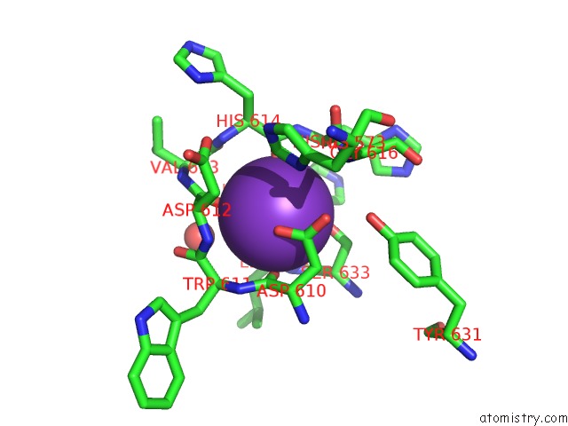
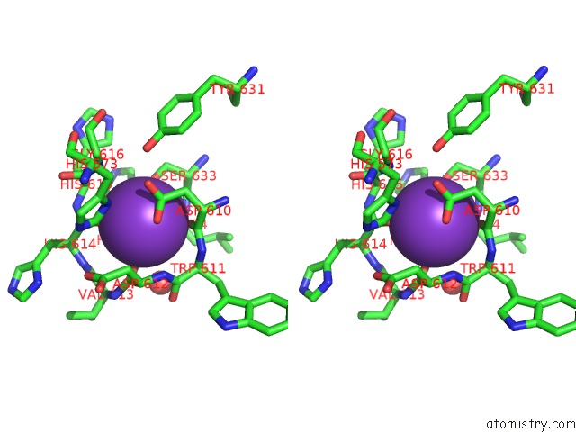
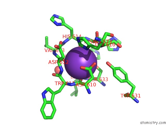
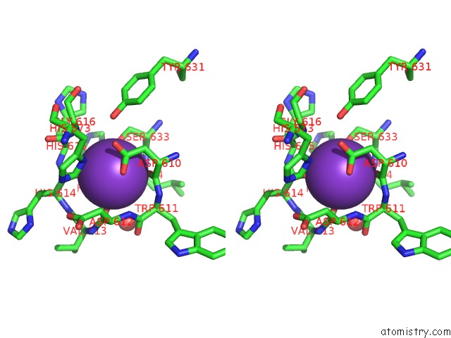
Potassium binding site 1 out of 6 in 5wgm
Go back to
Potassium binding site 1 out
of 6 in the Crystal Structure of Danio Rerio Histone Deacetylase 6 Catalytic Domain 2 in Complex with Acy-1083
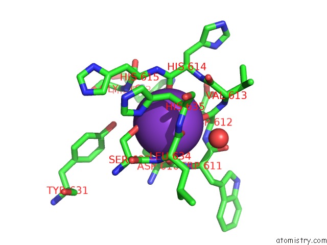
Mono view
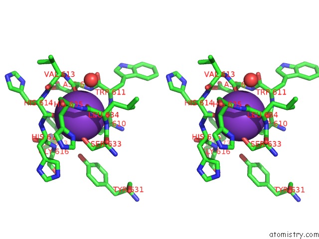
Stereo pair view
Mono view
Stereo pair view
A full contact list of Potassium with other atoms in the K binding
site number 1 of Crystal Structure of Danio Rerio Histone Deacetylase 6 Catalytic Domain 2 in Complex with Acy-1083 within 5.0Å range:
|
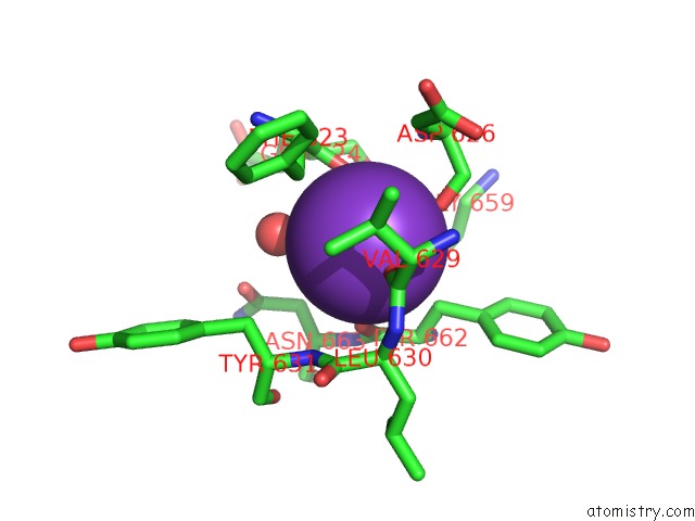
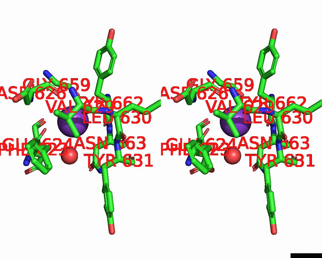
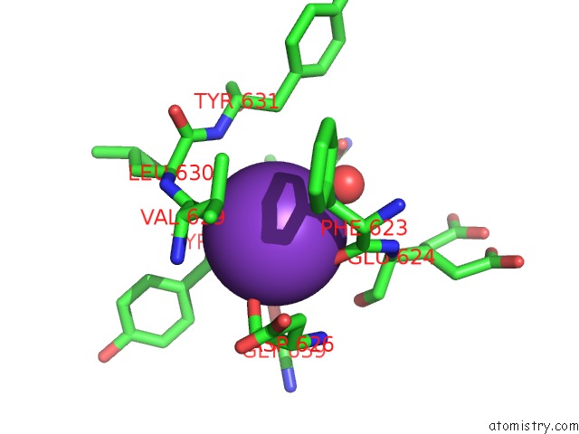
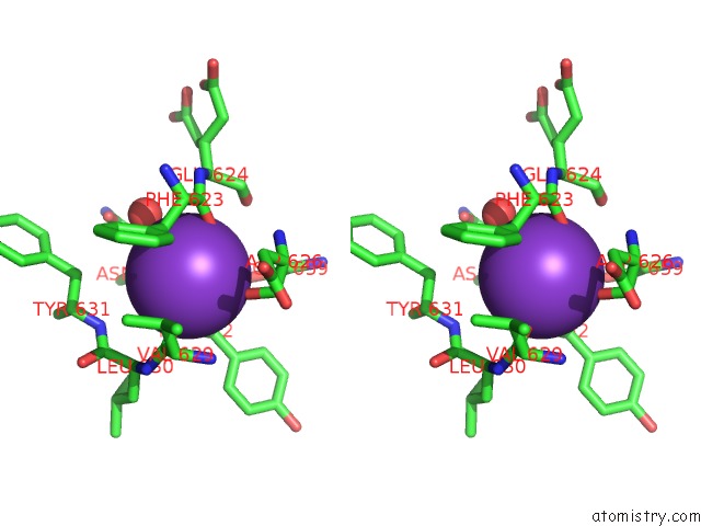
Potassium binding site 2 out of 6 in 5wgm
Go back to
Potassium binding site 2 out
of 6 in the Crystal Structure of Danio Rerio Histone Deacetylase 6 Catalytic Domain 2 in Complex with Acy-1083
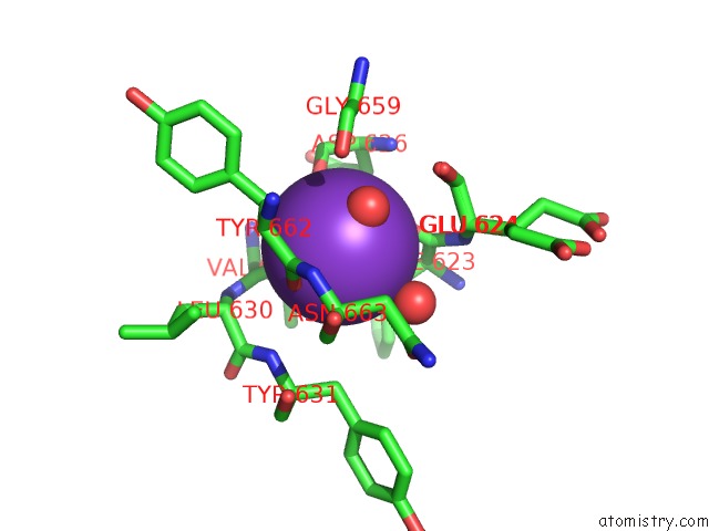
Mono view
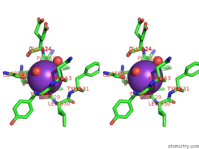
Stereo pair view
Mono view
Stereo pair view
A full contact list of Potassium with other atoms in the K binding
site number 2 of Crystal Structure of Danio Rerio Histone Deacetylase 6 Catalytic Domain 2 in Complex with Acy-1083 within 5.0Å range:
|
Potassium binding site 3 out of 6 in 5wgm
Go back to
Potassium binding site 3 out
of 6 in the Crystal Structure of Danio Rerio Histone Deacetylase 6 Catalytic Domain 2 in Complex with Acy-1083
Mono view
Stereo pair view
Mono view
Stereo pair view
A full contact list of Potassium with other atoms in the K binding
site number 3 of Crystal Structure of Danio Rerio Histone Deacetylase 6 Catalytic Domain 2 in Complex with Acy-1083 within 5.0Å range:
|
Potassium binding site 4 out of 6 in 5wgm
Go back to
Potassium binding site 4 out
of 6 in the Crystal Structure of Danio Rerio Histone Deacetylase 6 Catalytic Domain 2 in Complex with Acy-1083
Mono view
Stereo pair view
Mono view
Stereo pair view
A full contact list of Potassium with other atoms in the K binding
site number 4 of Crystal Structure of Danio Rerio Histone Deacetylase 6 Catalytic Domain 2 in Complex with Acy-1083 within 5.0Å range:
|
Potassium binding site 5 out of 6 in 5wgm
Go back to
Potassium binding site 5 out
of 6 in the Crystal Structure of Danio Rerio Histone Deacetylase 6 Catalytic Domain 2 in Complex with Acy-1083
Mono view
Stereo pair view
Mono view
Stereo pair view
A full contact list of Potassium with other atoms in the K binding
site number 5 of Crystal Structure of Danio Rerio Histone Deacetylase 6 Catalytic Domain 2 in Complex with Acy-1083 within 5.0Å range:
|
Potassium binding site 6 out of 6 in 5wgm
Go back to
Potassium binding site 6 out
of 6 in the Crystal Structure of Danio Rerio Histone Deacetylase 6 Catalytic Domain 2 in Complex with Acy-1083
Mono view
Stereo pair view
Mono view
Stereo pair view
A full contact list of Potassium with other atoms in the K binding
site number 6 of Crystal Structure of Danio Rerio Histone Deacetylase 6 Catalytic Domain 2 in Complex with Acy-1083 within 5.0Å range:
|
Reference:
N.J.Porter,
A.Mahendran,
R.Breslow,
D.W.Christianson.
Unusual Zinc-Binding Mode of HDAC6-Selective Hydroxamate Inhibitors. Proc. Natl. Acad. Sci. V. 114 13459 2017U.S.A..
ISSN: ESSN 1091-6490
PubMed: 29203661
DOI: 10.1073/PNAS.1718823114
Page generated: Sat Aug 9 10:04:37 2025
ISSN: ESSN 1091-6490
PubMed: 29203661
DOI: 10.1073/PNAS.1718823114
Last articles
Mg in 1HE1Mg in 1HDI
Mg in 1HCK
Mg in 1HBO
Mg in 1HBM
Mg in 1H8E
Mg in 1H8H
Mg in 1H7U
Mg in 1HA3
Mg in 1H7Q