Potassium »
PDB 5vt3-5x23 »
5w6h »
Potassium in PDB 5w6h: Crystal Structure of Bacteriophage CBA120 Tailspike Protein 4 Enzymatically Active Domain (TSP4DN, ORF213)
Protein crystallography data
The structure of Crystal Structure of Bacteriophage CBA120 Tailspike Protein 4 Enzymatically Active Domain (TSP4DN, ORF213), PDB code: 5w6h
was solved by
M.Plattner,
M.M.Shneider,
P.G.Leiman,
with X-Ray Crystallography technique. A brief refinement statistics is given in the table below:
| Resolution Low / High (Å) | 49.29 / 2.29 |
| Space group | P 41 2 2 |
| Cell size a, b, c (Å), α, β, γ (°) | 170.079, 170.079, 246.384, 90.00, 90.00, 90.00 |
| R / Rfree (%) | 14 / 17 |
Other elements in 5w6h:
The structure of Crystal Structure of Bacteriophage CBA120 Tailspike Protein 4 Enzymatically Active Domain (TSP4DN, ORF213) also contains other interesting chemical elements:
| Zinc | (Zn) | 1 atom |
| Chlorine | (Cl) | 1 atom |
| Sodium | (Na) | 1 atom |
Potassium Binding Sites:
The binding sites of Potassium atom in the Crystal Structure of Bacteriophage CBA120 Tailspike Protein 4 Enzymatically Active Domain (TSP4DN, ORF213)
(pdb code 5w6h). This binding sites where shown within
5.0 Angstroms radius around Potassium atom.
In total only one binding site of Potassium was determined in the Crystal Structure of Bacteriophage CBA120 Tailspike Protein 4 Enzymatically Active Domain (TSP4DN, ORF213), PDB code: 5w6h:
In total only one binding site of Potassium was determined in the Crystal Structure of Bacteriophage CBA120 Tailspike Protein 4 Enzymatically Active Domain (TSP4DN, ORF213), PDB code: 5w6h:
Potassium binding site 1 out of 1 in 5w6h
Go back to
Potassium binding site 1 out
of 1 in the Crystal Structure of Bacteriophage CBA120 Tailspike Protein 4 Enzymatically Active Domain (TSP4DN, ORF213)

Mono view
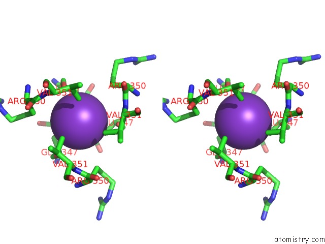
Stereo pair view
Mono view
Stereo pair view
A full contact list of Potassium with other atoms in the K binding
site number 1 of Crystal Structure of Bacteriophage CBA120 Tailspike Protein 4 Enzymatically Active Domain (TSP4DN, ORF213) within 5.0Å range:
|
Reference:
M.Plattner,
M.M.Shneider,
N.P.Arbatsky,
A.S.Shashkov,
A.O.Chizhov,
S.Nazarov,
N.S.Prokhorov,
N.M.I.Taylor,
S.A.Buth,
M.Gambino,
Y.E.Gencay,
L.Brondsted,
E.M.Kutter,
Y.A.Knirel,
P.G.Leiman.
Structure and Function of the Branched Receptor-Binding Complex of Bacteriophage CBA120. J.Mol.Biol. V. 431 3718 2019.
ISSN: ESSN 1089-8638
PubMed: 31325442
DOI: 10.1016/J.JMB.2019.07.022
Page generated: Mon Aug 12 14:53:12 2024
ISSN: ESSN 1089-8638
PubMed: 31325442
DOI: 10.1016/J.JMB.2019.07.022
Last articles
Zn in 9MJ5Zn in 9HNW
Zn in 9G0L
Zn in 9FNE
Zn in 9DZN
Zn in 9E0I
Zn in 9D32
Zn in 9DAK
Zn in 8ZXC
Zn in 8ZUF