Potassium »
PDB 5u3s-5vsn »
5vj9 »
Potassium in PDB 5vj9: Guanidine-II Riboswitch P2 Hairpin Dimer From Pseudomonas Aeruginosa
Protein crystallography data
The structure of Guanidine-II Riboswitch P2 Hairpin Dimer From Pseudomonas Aeruginosa, PDB code: 5vj9
was solved by
C.W.Reiss,
S.A.Strobel,
with X-Ray Crystallography technique. A brief refinement statistics is given in the table below:
| Resolution Low / High (Å) | 40.00 / 1.57 |
| Space group | P 21 21 21 |
| Cell size a, b, c (Å), α, β, γ (°) | 50.252, 60.664, 72.308, 90.00, 90.00, 90.00 |
| R / Rfree (%) | 19.2 / 23.4 |
Other elements in 5vj9:
The structure of Guanidine-II Riboswitch P2 Hairpin Dimer From Pseudomonas Aeruginosa also contains other interesting chemical elements:
| Magnesium | (Mg) | 1 atom |
Potassium Binding Sites:
The binding sites of Potassium atom in the Guanidine-II Riboswitch P2 Hairpin Dimer From Pseudomonas Aeruginosa
(pdb code 5vj9). This binding sites where shown within
5.0 Angstroms radius around Potassium atom.
In total only one binding site of Potassium was determined in the Guanidine-II Riboswitch P2 Hairpin Dimer From Pseudomonas Aeruginosa, PDB code: 5vj9:
In total only one binding site of Potassium was determined in the Guanidine-II Riboswitch P2 Hairpin Dimer From Pseudomonas Aeruginosa, PDB code: 5vj9:
Potassium binding site 1 out of 1 in 5vj9
Go back to
Potassium binding site 1 out
of 1 in the Guanidine-II Riboswitch P2 Hairpin Dimer From Pseudomonas Aeruginosa
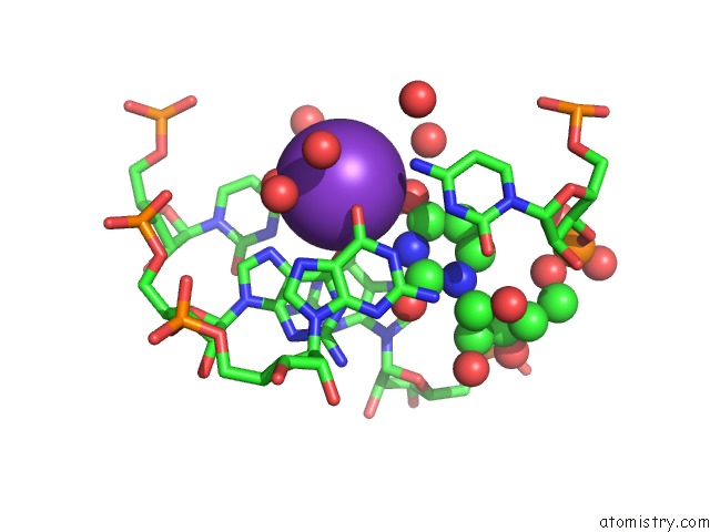
Mono view
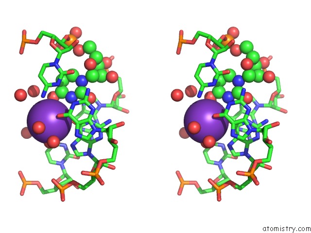
Stereo pair view
Mono view
Stereo pair view
A full contact list of Potassium with other atoms in the K binding
site number 1 of Guanidine-II Riboswitch P2 Hairpin Dimer From Pseudomonas Aeruginosa within 5.0Å range:
|
Reference:
C.W.Reiss,
S.A.Strobel.
Structural Basis For Ligand Binding to the Guanidine-II Riboswitch. Rna V. 23 1338 2017.
ISSN: ESSN 1469-9001
PubMed: 28600356
DOI: 10.1261/RNA.061804.117
Page generated: Sat Aug 9 09:59:41 2025
ISSN: ESSN 1469-9001
PubMed: 28600356
DOI: 10.1261/RNA.061804.117
Last articles
Mg in 1KK3Mg in 1KK2
Mg in 1KJY
Mg in 1KK1
Mg in 1KJJ
Mg in 1KJQ
Mg in 1KJI
Mg in 1KJ9
Mg in 1KJ8
Mg in 1KIZ