Potassium »
PDB 5mrm-5s9l »
5n2a »
Potassium in PDB 5n2a: Methyl-Coenzyme M Reductase III From Methanotorris Formicicus Trigonal Form
Enzymatic activity of Methyl-Coenzyme M Reductase III From Methanotorris Formicicus Trigonal Form
All present enzymatic activity of Methyl-Coenzyme M Reductase III From Methanotorris Formicicus Trigonal Form:
2.8.4.1;
2.8.4.1;
Protein crystallography data
The structure of Methyl-Coenzyme M Reductase III From Methanotorris Formicicus Trigonal Form, PDB code: 5n2a
was solved by
T.Wagner,
C.E.Wegner,
U.Ermler,
S.Shima,
with X-Ray Crystallography technique. A brief refinement statistics is given in the table below:
| Resolution Low / High (Å) | 39.85 / 2.80 |
| Space group | P 32 2 1 |
| Cell size a, b, c (Å), α, β, γ (°) | 127.699, 127.699, 160.376, 90.00, 90.00, 120.00 |
| R / Rfree (%) | 19.8 / 22.1 |
Other elements in 5n2a:
The structure of Methyl-Coenzyme M Reductase III From Methanotorris Formicicus Trigonal Form also contains other interesting chemical elements:
| Nickel | (Ni) | 1 atom |
| Bromine | (Br) | 1 atom |
Potassium Binding Sites:
The binding sites of Potassium atom in the Methyl-Coenzyme M Reductase III From Methanotorris Formicicus Trigonal Form
(pdb code 5n2a). This binding sites where shown within
5.0 Angstroms radius around Potassium atom.
In total only one binding site of Potassium was determined in the Methyl-Coenzyme M Reductase III From Methanotorris Formicicus Trigonal Form, PDB code: 5n2a:
In total only one binding site of Potassium was determined in the Methyl-Coenzyme M Reductase III From Methanotorris Formicicus Trigonal Form, PDB code: 5n2a:
Potassium binding site 1 out of 1 in 5n2a
Go back to
Potassium binding site 1 out
of 1 in the Methyl-Coenzyme M Reductase III From Methanotorris Formicicus Trigonal Form
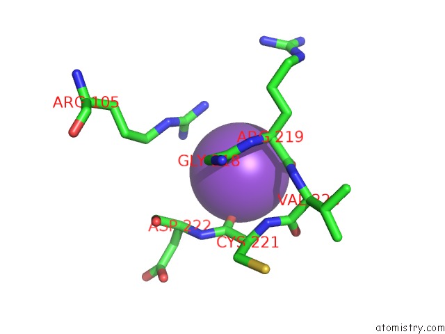
Mono view
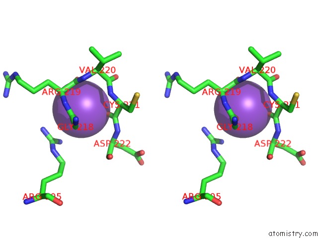
Stereo pair view
Mono view
Stereo pair view
A full contact list of Potassium with other atoms in the K binding
site number 1 of Methyl-Coenzyme M Reductase III From Methanotorris Formicicus Trigonal Form within 5.0Å range:
|
Reference:
T.Wagner,
C.E.Wegner,
J.Kahnt,
U.Ermler,
S.Shima.
Phylogenetic and Structural Comparisons of the Three Types of Methyl Coenzyme M Reductase From Methanococcales and Methanobacteriales. J.Bacteriol. V. 199 2017.
ISSN: ESSN 1098-5530
PubMed: 28559298
DOI: 10.1128/JB.00197-17
Page generated: Mon Aug 12 14:18:58 2024
ISSN: ESSN 1098-5530
PubMed: 28559298
DOI: 10.1128/JB.00197-17
Last articles
Zn in 9J0NZn in 9J0O
Zn in 9J0P
Zn in 9FJX
Zn in 9EKB
Zn in 9C0F
Zn in 9CAH
Zn in 9CH0
Zn in 9CH3
Zn in 9CH1