Potassium »
PDB 5mrm-5s9l »
5mrw »
Potassium in PDB 5mrw: Structure of the Kdpfabc Complex
Enzymatic activity of Structure of the Kdpfabc Complex
All present enzymatic activity of Structure of the Kdpfabc Complex:
3.6.3.12;
3.6.3.12;
Protein crystallography data
The structure of Structure of the Kdpfabc Complex, PDB code: 5mrw
was solved by
C.Huang,
B.P.Pedersen,
D.L.Stokes,
with X-Ray Crystallography technique. A brief refinement statistics is given in the table below:
| Resolution Low / High (Å) | 20.00 / 2.90 |
| Space group | P 1 21 1 |
| Cell size a, b, c (Å), α, β, γ (°) | 124.720, 166.290, 196.300, 90.00, 107.41, 90.00 |
| R / Rfree (%) | 24.3 / 27.5 |
Potassium Binding Sites:
The binding sites of Potassium atom in the Structure of the Kdpfabc Complex
(pdb code 5mrw). This binding sites where shown within
5.0 Angstroms radius around Potassium atom.
In total 3 binding sites of Potassium where determined in the Structure of the Kdpfabc Complex, PDB code: 5mrw:
Jump to Potassium binding site number: 1; 2; 3;
In total 3 binding sites of Potassium where determined in the Structure of the Kdpfabc Complex, PDB code: 5mrw:
Jump to Potassium binding site number: 1; 2; 3;
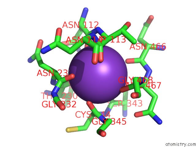
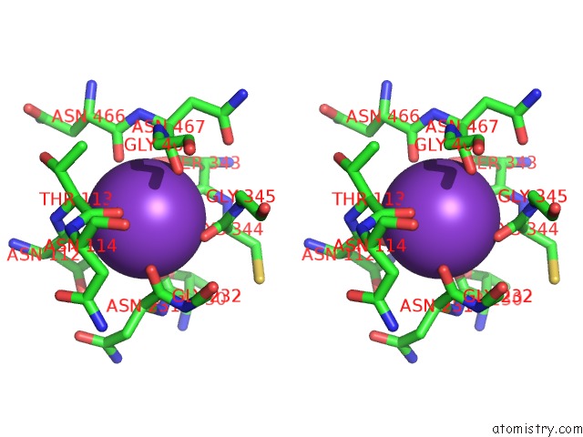
Potassium binding site 1 out of 3 in 5mrw
Go back to
Potassium binding site 1 out
of 3 in the Structure of the Kdpfabc Complex
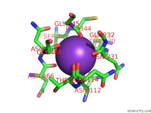
Mono view
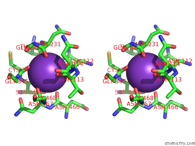
Stereo pair view
Mono view
Stereo pair view
A full contact list of Potassium with other atoms in the K binding
site number 1 of Structure of the Kdpfabc Complex within 5.0Å range:
|
Potassium binding site 2 out of 3 in 5mrw
Go back to
Potassium binding site 2 out
of 3 in the Structure of the Kdpfabc Complex
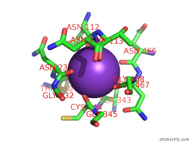
Mono view
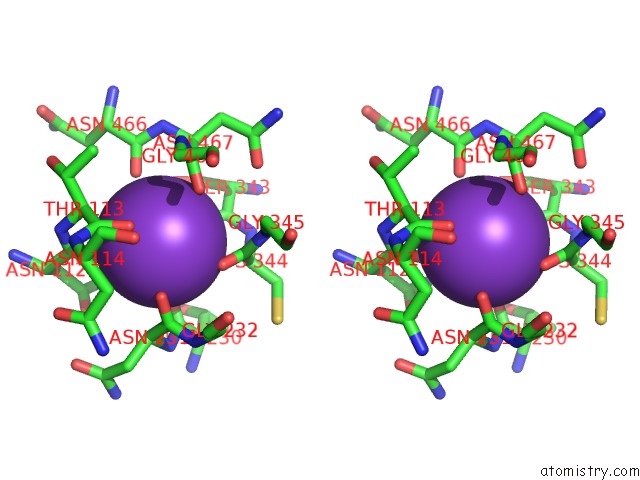
Stereo pair view
Mono view
Stereo pair view
A full contact list of Potassium with other atoms in the K binding
site number 2 of Structure of the Kdpfabc Complex within 5.0Å range:
|
Potassium binding site 3 out of 3 in 5mrw
Go back to
Potassium binding site 3 out
of 3 in the Structure of the Kdpfabc Complex
Mono view
Stereo pair view
Mono view
Stereo pair view
A full contact list of Potassium with other atoms in the K binding
site number 3 of Structure of the Kdpfabc Complex within 5.0Å range:
|
Reference:
C.S.Huang,
B.P.Pedersen,
D.L.Stokes.
Crystal Structure of the Potassium-Importing Kdpfabc Membrane Complex. Nature V. 546 681 2017.
ISSN: ESSN 1476-4687
PubMed: 28636601
DOI: 10.1038/NATURE22970
Page generated: Mon Aug 12 14:18:26 2024
ISSN: ESSN 1476-4687
PubMed: 28636601
DOI: 10.1038/NATURE22970
Last articles
Zn in 9J0NZn in 9J0O
Zn in 9J0P
Zn in 9FJX
Zn in 9EKB
Zn in 9C0F
Zn in 9CAH
Zn in 9CH0
Zn in 9CH3
Zn in 9CH1