Potassium »
PDB 5ksd-5mq0 »
5lk1 »
Potassium in PDB 5lk1: Structure of Hantavirus Envelope Glycoprotein Gc in Postfusion Conformation in Presence of 200 Mm Kcl
Protein crystallography data
The structure of Structure of Hantavirus Envelope Glycoprotein Gc in Postfusion Conformation in Presence of 200 Mm Kcl, PDB code: 5lk1
was solved by
P.Guardado-Calvo,
F.A.Rey,
with X-Ray Crystallography technique. A brief refinement statistics is given in the table below:
| Resolution Low / High (Å) | 37.55 / 1.70 |
| Space group | H 3 |
| Cell size a, b, c (Å), α, β, γ (°) | 107.289, 107.289, 127.539, 90.00, 90.00, 120.00 |
| R / Rfree (%) | 17.7 / 20.3 |
Other elements in 5lk1:
The structure of Structure of Hantavirus Envelope Glycoprotein Gc in Postfusion Conformation in Presence of 200 Mm Kcl also contains other interesting chemical elements:
| Sodium | (Na) | 1 atom |
Potassium Binding Sites:
The binding sites of Potassium atom in the Structure of Hantavirus Envelope Glycoprotein Gc in Postfusion Conformation in Presence of 200 Mm Kcl
(pdb code 5lk1). This binding sites where shown within
5.0 Angstroms radius around Potassium atom.
In total only one binding site of Potassium was determined in the Structure of Hantavirus Envelope Glycoprotein Gc in Postfusion Conformation in Presence of 200 Mm Kcl, PDB code: 5lk1:
In total only one binding site of Potassium was determined in the Structure of Hantavirus Envelope Glycoprotein Gc in Postfusion Conformation in Presence of 200 Mm Kcl, PDB code: 5lk1:
Potassium binding site 1 out of 1 in 5lk1
Go back to
Potassium binding site 1 out
of 1 in the Structure of Hantavirus Envelope Glycoprotein Gc in Postfusion Conformation in Presence of 200 Mm Kcl
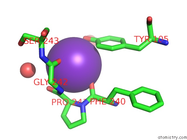
Mono view
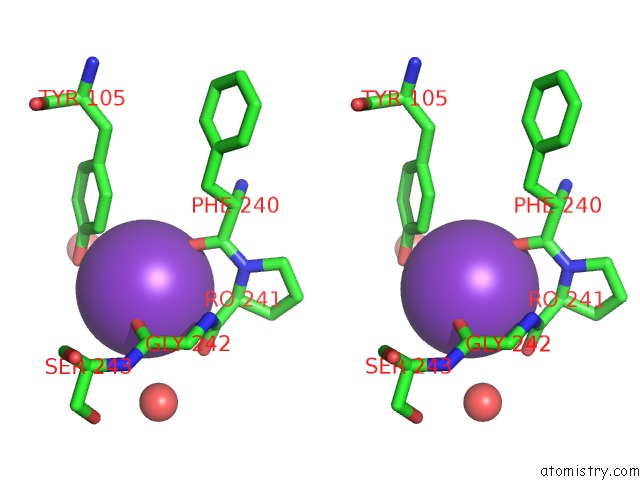
Stereo pair view
Mono view
Stereo pair view
A full contact list of Potassium with other atoms in the K binding
site number 1 of Structure of Hantavirus Envelope Glycoprotein Gc in Postfusion Conformation in Presence of 200 Mm Kcl within 5.0Å range:
|
Reference:
P.Guardado-Calvo,
E.A.Bignon,
E.Stettner,
S.A.Jeffers,
J.Perez-Vargas,
G.Pehau-Arnaudet,
M.A.Tortorici,
J.L.Jestin,
P.England,
N.D.Tischler,
F.A.Rey.
Mechanistic Insight Into Bunyavirus-Induced Membrane Fusion From Structure-Function Analyses of the Hantavirus Envelope Glycoprotein Gc. Plos Pathog. V. 12 05813 2016.
ISSN: ESSN 1553-7374
PubMed: 27783711
DOI: 10.1371/JOURNAL.PPAT.1005813
Page generated: Mon Aug 12 14:14:18 2024
ISSN: ESSN 1553-7374
PubMed: 27783711
DOI: 10.1371/JOURNAL.PPAT.1005813
Last articles
Zn in 9J0NZn in 9J0O
Zn in 9J0P
Zn in 9FJX
Zn in 9EKB
Zn in 9C0F
Zn in 9CAH
Zn in 9CH0
Zn in 9CH3
Zn in 9CH1