Potassium »
PDB 5irf-5koe »
5k03 »
Potassium in PDB 5k03: Crystal Structure of Comt in Complex with 2,6-Dimethyl-3-(1H-Pyrazol- 3-Yl)Imidazo[1,2-A]Pyridine
Enzymatic activity of Crystal Structure of Comt in Complex with 2,6-Dimethyl-3-(1H-Pyrazol- 3-Yl)Imidazo[1,2-A]Pyridine
All present enzymatic activity of Crystal Structure of Comt in Complex with 2,6-Dimethyl-3-(1H-Pyrazol- 3-Yl)Imidazo[1,2-A]Pyridine:
2.1.1.6;
2.1.1.6;
Protein crystallography data
The structure of Crystal Structure of Comt in Complex with 2,6-Dimethyl-3-(1H-Pyrazol- 3-Yl)Imidazo[1,2-A]Pyridine, PDB code: 5k03
was solved by
A.Ehler,
R.M.Rodriguez-Sarmiento,
M.G.Rudolph,
with X-Ray Crystallography technique. A brief refinement statistics is given in the table below:
| Resolution Low / High (Å) | 30.49 / 1.81 |
| Space group | P 21 21 21 |
| Cell size a, b, c (Å), α, β, γ (°) | 33.357, 59.670, 106.395, 90.00, 90.00, 90.00 |
| R / Rfree (%) | 19.9 / 25.3 |
Potassium Binding Sites:
The binding sites of Potassium atom in the Crystal Structure of Comt in Complex with 2,6-Dimethyl-3-(1H-Pyrazol- 3-Yl)Imidazo[1,2-A]Pyridine
(pdb code 5k03). This binding sites where shown within
5.0 Angstroms radius around Potassium atom.
In total only one binding site of Potassium was determined in the Crystal Structure of Comt in Complex with 2,6-Dimethyl-3-(1H-Pyrazol- 3-Yl)Imidazo[1,2-A]Pyridine, PDB code: 5k03:
In total only one binding site of Potassium was determined in the Crystal Structure of Comt in Complex with 2,6-Dimethyl-3-(1H-Pyrazol- 3-Yl)Imidazo[1,2-A]Pyridine, PDB code: 5k03:
Potassium binding site 1 out of 1 in 5k03
Go back to
Potassium binding site 1 out
of 1 in the Crystal Structure of Comt in Complex with 2,6-Dimethyl-3-(1H-Pyrazol- 3-Yl)Imidazo[1,2-A]Pyridine
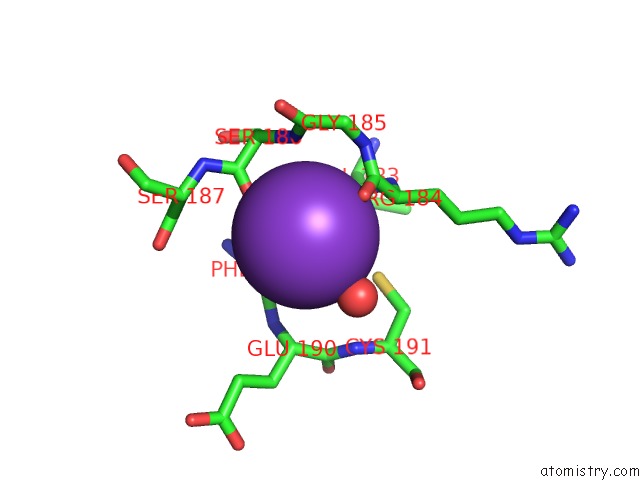
Mono view
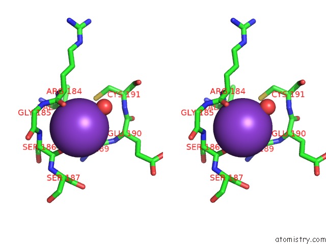
Stereo pair view
Mono view
Stereo pair view
A full contact list of Potassium with other atoms in the K binding
site number 1 of Crystal Structure of Comt in Complex with 2,6-Dimethyl-3-(1H-Pyrazol- 3-Yl)Imidazo[1,2-A]Pyridine within 5.0Å range:
|
Reference:
C.Lerner,
R.Jakob-Roetne,
B.Buettelmann,
A.Ehler,
M.Rudolph,
R.M.Rodriguez Sarmiento.
Design of Potent and Druglike Nonphenolic Inhibitors For Catechol O-Methyltransferase Derived From A Fragment Screening Approach Targeting the S-Adenosyl-L-Methionine Pocket. J. Med. Chem. V. 59 10163 2016.
ISSN: ISSN 1520-4804
PubMed: 27685665
DOI: 10.1021/ACS.JMEDCHEM.6B00927
Page generated: Mon Aug 12 13:57:55 2024
ISSN: ISSN 1520-4804
PubMed: 27685665
DOI: 10.1021/ACS.JMEDCHEM.6B00927
Last articles
Zn in 9J0NZn in 9J0O
Zn in 9J0P
Zn in 9FJX
Zn in 9EKB
Zn in 9C0F
Zn in 9CAH
Zn in 9CH0
Zn in 9CH3
Zn in 9CH1