Potassium »
PDB 5g17-5imu »
5imu »
Potassium in PDB 5imu: A Fragment of Conserved Hypothetical Protein RV3899C (Residues 184- 410) From Mycobacterium Tuberculosis
Protein crystallography data
The structure of A Fragment of Conserved Hypothetical Protein RV3899C (Residues 184- 410) From Mycobacterium Tuberculosis, PDB code: 5imu
was solved by
D.F.Li,
Y.R.Gao,
Y.Y.Liu,
L.J.Bi,
with X-Ray Crystallography technique. A brief refinement statistics is given in the table below:
| Resolution Low / High (Å) | 44.50 / 1.90 |
| Space group | P 21 21 21 |
| Cell size a, b, c (Å), α, β, γ (°) | 50.155, 54.973, 75.807, 90.00, 90.00, 90.00 |
| R / Rfree (%) | 16 / 19 |
Potassium Binding Sites:
The binding sites of Potassium atom in the A Fragment of Conserved Hypothetical Protein RV3899C (Residues 184- 410) From Mycobacterium Tuberculosis
(pdb code 5imu). This binding sites where shown within
5.0 Angstroms radius around Potassium atom.
In total only one binding site of Potassium was determined in the A Fragment of Conserved Hypothetical Protein RV3899C (Residues 184- 410) From Mycobacterium Tuberculosis, PDB code: 5imu:
In total only one binding site of Potassium was determined in the A Fragment of Conserved Hypothetical Protein RV3899C (Residues 184- 410) From Mycobacterium Tuberculosis, PDB code: 5imu:
Potassium binding site 1 out of 1 in 5imu
Go back to
Potassium binding site 1 out
of 1 in the A Fragment of Conserved Hypothetical Protein RV3899C (Residues 184- 410) From Mycobacterium Tuberculosis
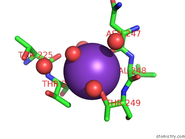
Mono view
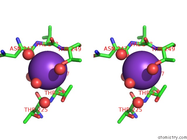
Stereo pair view
Mono view
Stereo pair view
A full contact list of Potassium with other atoms in the K binding
site number 1 of A Fragment of Conserved Hypothetical Protein RV3899C (Residues 184- 410) From Mycobacterium Tuberculosis within 5.0Å range:
|
Reference:
Y.Y.Liu,
Y.R.Gao,
D.F.Li,
J.Fleming,
H.L.Li,
L.J.Bi.
Crystal Structure of RV3899C(184-410), A Hypothetical Protein From Mycobacterium Tuberculosis Acta Crystallogr F Struct V. 72 642 2016BIOL Commun.
ISSN: ESSN 2053-230X
PubMed: 27487929
DOI: 10.1107/S2053230X16010943
Page generated: Mon Aug 12 13:55:06 2024
ISSN: ESSN 2053-230X
PubMed: 27487929
DOI: 10.1107/S2053230X16010943
Last articles
Zn in 9J0NZn in 9J0O
Zn in 9J0P
Zn in 9FJX
Zn in 9EKB
Zn in 9C0F
Zn in 9CAH
Zn in 9CH0
Zn in 9CH3
Zn in 9CH1