Potassium »
PDB 5g17-5imu »
5h6g »
Potassium in PDB 5h6g: Crystal Structure of A Thermostable Lipase From Marine Streptomyces
Protein crystallography data
The structure of Crystal Structure of A Thermostable Lipase From Marine Streptomyces, PDB code: 5h6g
was solved by
S.Hou,
Z.Zhao,
J.Liu,
with X-Ray Crystallography technique. A brief refinement statistics is given in the table below:
| Resolution Low / High (Å) | 64.76 / 2.34 |
| Space group | P 32 2 1 |
| Cell size a, b, c (Å), α, β, γ (°) | 129.500, 129.500, 137.830, 90.00, 90.00, 120.00 |
| R / Rfree (%) | 19.1 / 22.2 |
Other elements in 5h6g:
The structure of Crystal Structure of A Thermostable Lipase From Marine Streptomyces also contains other interesting chemical elements:
| Chlorine | (Cl) | 2 atoms |
Potassium Binding Sites:
The binding sites of Potassium atom in the Crystal Structure of A Thermostable Lipase From Marine Streptomyces
(pdb code 5h6g). This binding sites where shown within
5.0 Angstroms radius around Potassium atom.
In total only one binding site of Potassium was determined in the Crystal Structure of A Thermostable Lipase From Marine Streptomyces, PDB code: 5h6g:
In total only one binding site of Potassium was determined in the Crystal Structure of A Thermostable Lipase From Marine Streptomyces, PDB code: 5h6g:
Potassium binding site 1 out of 1 in 5h6g
Go back to
Potassium binding site 1 out
of 1 in the Crystal Structure of A Thermostable Lipase From Marine Streptomyces

Mono view
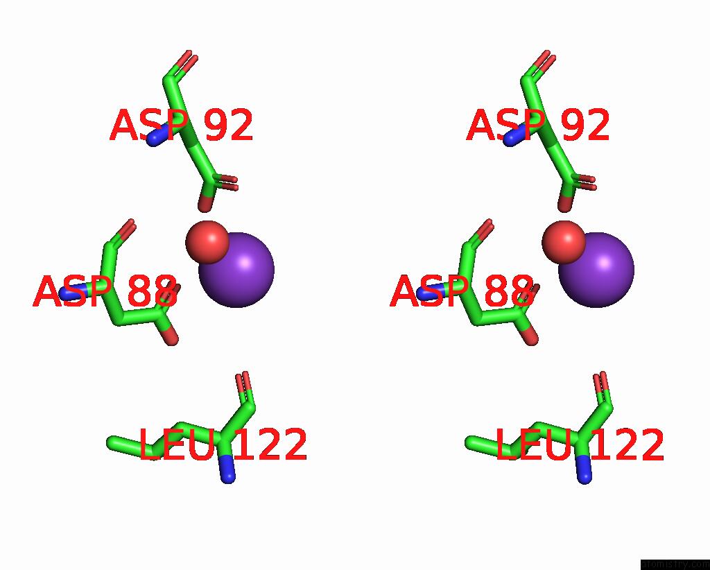
Stereo pair view
Mono view
Stereo pair view
A full contact list of Potassium with other atoms in the K binding
site number 1 of Crystal Structure of A Thermostable Lipase From Marine Streptomyces within 5.0Å range:
|
Reference:
Z.Zhao,
S.Hou,
D.Lan,
X.Wang,
J.Liu,
F.I.Khan,
Y.Wang.
Crystal Structure of A Lipase From Streptomyces Sp. Strain W007 - Implications For Thermostability and Regiospecificity Febs J. V. 284 3506 2017.
ISSN: ISSN 1742-4658
PubMed: 28857479
DOI: 10.1111/FEBS.14211
Page generated: Mon Aug 12 13:47:55 2024
ISSN: ISSN 1742-4658
PubMed: 28857479
DOI: 10.1111/FEBS.14211
Last articles
Zn in 9J0NZn in 9J0O
Zn in 9J0P
Zn in 9FJX
Zn in 9EKB
Zn in 9C0F
Zn in 9CAH
Zn in 9CH0
Zn in 9CH3
Zn in 9CH1