Potassium in PDB 5fcw: HDAC8 Complexed with A Hydroxamic Acid
Enzymatic activity of HDAC8 Complexed with A Hydroxamic Acid
All present enzymatic activity of HDAC8 Complexed with A Hydroxamic Acid:
3.5.1.98;
Protein crystallography data
The structure of HDAC8 Complexed with A Hydroxamic Acid, PDB code: 5fcw
was solved by
K.E.Cole,
K.Perry,
with X-Ray Crystallography technique. A brief refinement statistics is given in the table below:
Resolution Low / High (Å)
|
49.66 /
1.98
|
Space group
|
P 1 21 1
|
Cell size a, b, c (Å), α, β, γ (°)
|
53.443,
84.548,
94.307,
90.00,
100.08,
90.00
|
R / Rfree (%)
|
17.2 /
21.6
|
Other elements in 5fcw:
The structure of HDAC8 Complexed with A Hydroxamic Acid also contains other interesting chemical elements:
Potassium Binding Sites:
The binding sites of Potassium atom in the HDAC8 Complexed with A Hydroxamic Acid
(pdb code 5fcw). This binding sites where shown within
5.0 Angstroms radius around Potassium atom.
In total 2 binding sites of Potassium where determined in the
HDAC8 Complexed with A Hydroxamic Acid, PDB code: 5fcw:
Jump to Potassium binding site number:
1;
2;
Potassium binding site 1 out
of 2 in 5fcw
Go back to
Potassium Binding Sites List in 5fcw
Potassium binding site 1 out
of 2 in the HDAC8 Complexed with A Hydroxamic Acid
 Mono view
 Stereo pair view
|
A full contact list of Potassium with other atoms in the K binding
site number 1 of HDAC8 Complexed with A Hydroxamic Acid within 5.0Å range:
probe
|
atom
|
residue
|
distance (Å)
|
B
|
Occ
|
A:K403
b:14.0
occ:1.00
|
O
|
A:LEU200
|
2.6
|
14.5
|
1.0
|
OD1
|
A:ASP176
|
2.7
|
14.0
|
1.0
|
O
|
A:ASP178
|
2.7
|
15.7
|
1.0
|
O
|
A:ASP176
|
2.7
|
11.8
|
1.0
|
O
|
A:HIS180
|
2.8
|
12.6
|
1.0
|
OG
|
A:SER199
|
2.9
|
16.6
|
1.0
|
C
|
A:ASP176
|
3.4
|
15.6
|
1.0
|
CG
|
A:ASP176
|
3.4
|
17.8
|
1.0
|
N
|
A:ASP178
|
3.5
|
14.9
|
1.0
|
C
|
A:ASP178
|
3.6
|
14.7
|
1.0
|
C
|
A:LEU200
|
3.6
|
16.6
|
1.0
|
CB
|
A:ASP176
|
3.7
|
14.9
|
1.0
|
CB
|
A:HIS201
|
3.8
|
12.5
|
1.0
|
C
|
A:HIS180
|
3.8
|
12.7
|
1.0
|
CA
|
A:ASP178
|
3.9
|
14.9
|
1.0
|
N
|
A:LEU200
|
3.9
|
15.7
|
1.0
|
C
|
A:LEU177
|
3.9
|
14.6
|
1.0
|
N
|
A:LEU177
|
4.0
|
14.3
|
1.0
|
CB
|
A:ASP178
|
4.0
|
14.3
|
1.0
|
CB
|
A:SER199
|
4.1
|
13.4
|
1.0
|
CA
|
A:LEU177
|
4.2
|
13.7
|
1.0
|
CA
|
A:ASP176
|
4.2
|
15.3
|
1.0
|
CA
|
A:SER199
|
4.3
|
16.1
|
1.0
|
CA
|
A:HIS201
|
4.3
|
16.5
|
1.0
|
OD2
|
A:ASP176
|
4.3
|
19.2
|
1.0
|
N
|
A:HIS201
|
4.4
|
15.4
|
1.0
|
N
|
A:GLY182
|
4.4
|
13.6
|
1.0
|
C
|
A:SER199
|
4.4
|
17.1
|
1.0
|
CA
|
A:HIS181
|
4.5
|
14.1
|
1.0
|
CA
|
A:LEU200
|
4.5
|
16.4
|
1.0
|
N
|
A:HIS180
|
4.5
|
14.5
|
1.0
|
ND1
|
A:HIS201
|
4.5
|
14.3
|
1.0
|
O
|
A:HOH529
|
4.5
|
15.9
|
1.0
|
N
|
A:HIS181
|
4.5
|
12.1
|
1.0
|
CG
|
A:HIS201
|
4.6
|
14.5
|
1.0
|
O
|
A:LEU177
|
4.7
|
13.3
|
1.0
|
C
|
A:LEU179
|
4.7
|
15.7
|
1.0
|
N
|
A:LEU179
|
4.7
|
13.8
|
1.0
|
CA
|
A:HIS180
|
4.8
|
11.4
|
1.0
|
C
|
A:HIS181
|
4.8
|
17.3
|
1.0
|
CE1
|
A:HIS142
|
4.9
|
14.9
|
1.0
|
|
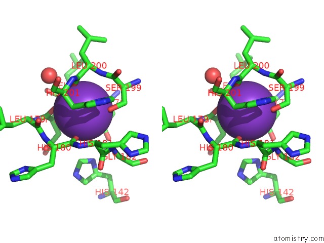
Potassium binding site 2 out
of 2 in 5fcw
Go back to
Potassium Binding Sites List in 5fcw
Potassium binding site 2 out
of 2 in the HDAC8 Complexed with A Hydroxamic Acid
 Mono view
 Stereo pair view
|
A full contact list of Potassium with other atoms in the K binding
site number 2 of HDAC8 Complexed with A Hydroxamic Acid within 5.0Å range:
probe
|
atom
|
residue
|
distance (Å)
|
B
|
Occ
|
B:K403
b:16.2
occ:1.00
|
O
|
B:ASP178
|
2.5
|
14.5
|
1.0
|
O
|
B:LEU200
|
2.6
|
16.8
|
1.0
|
OD1
|
B:ASP176
|
2.7
|
21.0
|
1.0
|
O
|
B:HIS180
|
2.7
|
13.6
|
1.0
|
O
|
B:ASP176
|
2.7
|
21.5
|
1.0
|
OG
|
B:SER199
|
2.9
|
19.5
|
1.0
|
CG
|
B:ASP176
|
3.4
|
19.9
|
1.0
|
C
|
B:ASP176
|
3.5
|
19.9
|
1.0
|
C
|
B:ASP178
|
3.5
|
19.9
|
1.0
|
N
|
B:ASP178
|
3.5
|
19.3
|
1.0
|
C
|
B:LEU200
|
3.6
|
21.4
|
1.0
|
C
|
B:HIS180
|
3.7
|
15.3
|
1.0
|
CB
|
B:HIS201
|
3.8
|
14.2
|
1.0
|
CB
|
B:ASP176
|
3.8
|
15.0
|
1.0
|
N
|
B:LEU200
|
3.9
|
19.0
|
1.0
|
CA
|
B:ASP178
|
3.9
|
17.4
|
1.0
|
C
|
B:LEU177
|
4.0
|
18.9
|
1.0
|
CB
|
B:ASP178
|
4.1
|
20.2
|
1.0
|
CB
|
B:SER199
|
4.1
|
19.2
|
1.0
|
N
|
B:LEU177
|
4.1
|
16.4
|
1.0
|
CA
|
B:LEU177
|
4.3
|
14.9
|
1.0
|
CA
|
B:ASP176
|
4.3
|
19.0
|
1.0
|
CA
|
B:HIS201
|
4.3
|
17.1
|
1.0
|
ND1
|
B:HIS201
|
4.3
|
17.1
|
1.0
|
CA
|
B:SER199
|
4.3
|
23.7
|
1.0
|
OD2
|
B:ASP176
|
4.3
|
20.7
|
1.0
|
N
|
B:HIS180
|
4.4
|
18.0
|
1.0
|
N
|
B:GLY182
|
4.4
|
17.2
|
1.0
|
N
|
B:HIS201
|
4.4
|
19.0
|
1.0
|
C
|
B:SER199
|
4.4
|
22.6
|
1.0
|
CA
|
B:LEU200
|
4.4
|
18.1
|
1.0
|
CA
|
B:HIS181
|
4.5
|
15.1
|
1.0
|
N
|
B:HIS181
|
4.5
|
13.3
|
1.0
|
O
|
B:HOH528
|
4.5
|
12.2
|
1.0
|
CG
|
B:HIS201
|
4.5
|
13.4
|
1.0
|
C
|
B:LEU179
|
4.6
|
18.9
|
1.0
|
N
|
B:LEU179
|
4.6
|
17.8
|
1.0
|
CA
|
B:HIS180
|
4.7
|
16.5
|
1.0
|
O
|
B:LEU177
|
4.8
|
16.5
|
1.0
|
C
|
B:HIS181
|
4.8
|
16.3
|
1.0
|
CE1
|
B:HIS142
|
4.9
|
20.7
|
1.0
|
O
|
B:LEU179
|
4.9
|
16.6
|
1.0
|
|
Reference:
A.A.Tabackman,
R.Frankson,
E.S.Marsan,
K.Perry,
K.E.Cole.
Structure of 'Linkerless' Hydroxamic Acid Inhibitor-HDAC8 Complex Confirms the Formation of An Isoform-Specific Subpocket. J.Struct.Biol. V. 195 373 2016.
ISSN: ESSN 1095-8657
PubMed: 27374062
DOI: 10.1016/J.JSB.2016.06.023
Page generated: Mon Aug 12 13:31:28 2024
|