Potassium »
PDB 5dkp-5f0g »
5eek »
Potassium in PDB 5eek: Crystal Structure of Danio Rerio Histone Deacetylase 6 Catalytic Domain 2 in Complex with Trichostatin A
Enzymatic activity of Crystal Structure of Danio Rerio Histone Deacetylase 6 Catalytic Domain 2 in Complex with Trichostatin A
All present enzymatic activity of Crystal Structure of Danio Rerio Histone Deacetylase 6 Catalytic Domain 2 in Complex with Trichostatin A:
3.5.1.98;
3.5.1.98;
Protein crystallography data
The structure of Crystal Structure of Danio Rerio Histone Deacetylase 6 Catalytic Domain 2 in Complex with Trichostatin A, PDB code: 5eek
was solved by
Y.Hai,
D.W.Christianson,
with X-Ray Crystallography technique. A brief refinement statistics is given in the table below:
| Resolution Low / High (Å) | 44.01 / 1.59 |
| Space group | P 21 21 2 |
| Cell size a, b, c (Å), α, β, γ (°) | 83.881, 94.429, 51.698, 90.00, 90.00, 90.00 |
| R / Rfree (%) | 13 / 16.3 |
Other elements in 5eek:
The structure of Crystal Structure of Danio Rerio Histone Deacetylase 6 Catalytic Domain 2 in Complex with Trichostatin A also contains other interesting chemical elements:
| Zinc | (Zn) | 1 atom |
| Iodine | (I) | 31 atoms |
| Chlorine | (Cl) | 1 atom |
Potassium Binding Sites:
The binding sites of Potassium atom in the Crystal Structure of Danio Rerio Histone Deacetylase 6 Catalytic Domain 2 in Complex with Trichostatin A
(pdb code 5eek). This binding sites where shown within
5.0 Angstroms radius around Potassium atom.
In total 2 binding sites of Potassium where determined in the Crystal Structure of Danio Rerio Histone Deacetylase 6 Catalytic Domain 2 in Complex with Trichostatin A, PDB code: 5eek:
Jump to Potassium binding site number: 1; 2;
In total 2 binding sites of Potassium where determined in the Crystal Structure of Danio Rerio Histone Deacetylase 6 Catalytic Domain 2 in Complex with Trichostatin A, PDB code: 5eek:
Jump to Potassium binding site number: 1; 2;
Potassium binding site 1 out of 2 in 5eek
Go back to
Potassium binding site 1 out
of 2 in the Crystal Structure of Danio Rerio Histone Deacetylase 6 Catalytic Domain 2 in Complex with Trichostatin A
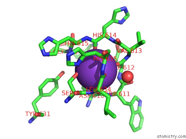
Mono view
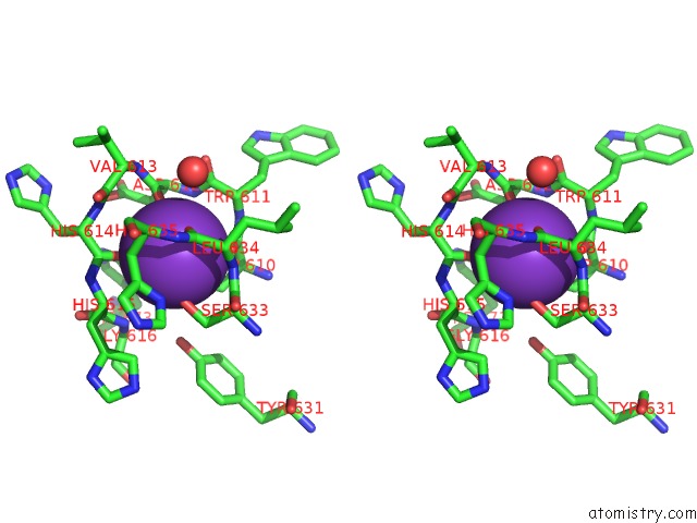
Stereo pair view
Mono view
Stereo pair view
A full contact list of Potassium with other atoms in the K binding
site number 1 of Crystal Structure of Danio Rerio Histone Deacetylase 6 Catalytic Domain 2 in Complex with Trichostatin A within 5.0Å range:
|
Potassium binding site 2 out of 2 in 5eek
Go back to
Potassium binding site 2 out
of 2 in the Crystal Structure of Danio Rerio Histone Deacetylase 6 Catalytic Domain 2 in Complex with Trichostatin A
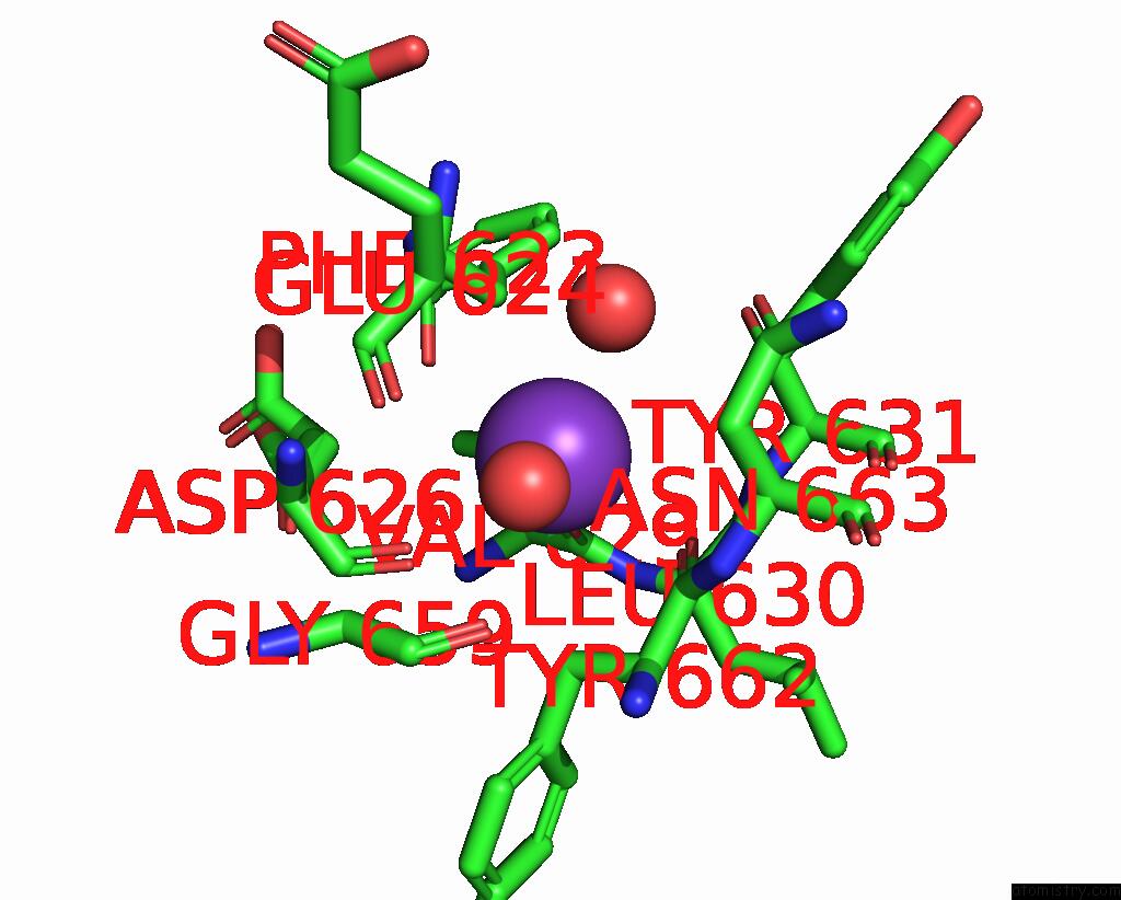
Mono view
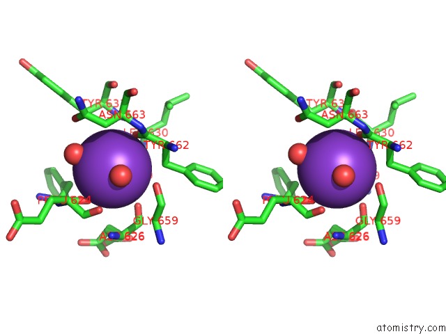
Stereo pair view
Mono view
Stereo pair view
A full contact list of Potassium with other atoms in the K binding
site number 2 of Crystal Structure of Danio Rerio Histone Deacetylase 6 Catalytic Domain 2 in Complex with Trichostatin A within 5.0Å range:
|
Reference:
Y.Hai,
D.W.Christianson.
Histone Deacetylase 6 Structure and Molecular Basis of Catalysis and Inhibition. Nat.Chem.Biol. V. 12 741 2016.
ISSN: ESSN 1552-4469
PubMed: 27454933
DOI: 10.1038/NCHEMBIO.2134
Page generated: Mon Aug 12 13:24:07 2024
ISSN: ESSN 1552-4469
PubMed: 27454933
DOI: 10.1038/NCHEMBIO.2134
Last articles
Zn in 9J0NZn in 9J0O
Zn in 9J0P
Zn in 9FJX
Zn in 9EKB
Zn in 9C0F
Zn in 9CAH
Zn in 9CH0
Zn in 9CH3
Zn in 9CH1