Potassium »
PDB 5dkp-5f0g »
5dou »
Potassium in PDB 5dou: Crystal Structure of Human Carbamoyl Phosphate Synthetase I (CPS1), Ligand-Bound Form
Enzymatic activity of Crystal Structure of Human Carbamoyl Phosphate Synthetase I (CPS1), Ligand-Bound Form
All present enzymatic activity of Crystal Structure of Human Carbamoyl Phosphate Synthetase I (CPS1), Ligand-Bound Form:
6.3.4.16;
6.3.4.16;
Protein crystallography data
The structure of Crystal Structure of Human Carbamoyl Phosphate Synthetase I (CPS1), Ligand-Bound Form, PDB code: 5dou
was solved by
S.De Cima,
L.M.Polo,
I.Fita,
V.Rubio,
with X-Ray Crystallography technique. A brief refinement statistics is given in the table below:
| Resolution Low / High (Å) | 40.00 / 2.60 |
| Space group | P 1 |
| Cell size a, b, c (Å), α, β, γ (°) | 78.919, 98.558, 214.890, 90.66, 98.65, 90.08 |
| R / Rfree (%) | 19.5 / 22.9 |
Other elements in 5dou:
The structure of Crystal Structure of Human Carbamoyl Phosphate Synthetase I (CPS1), Ligand-Bound Form also contains other interesting chemical elements:
| Nickel | (Ni) | 4 atoms |
| Magnesium | (Mg) | 12 atoms |
| Chlorine | (Cl) | 3 atoms |
Potassium Binding Sites:
Pages:
>>> Page 1 <<< Page 2, Binding sites: 11 - 18;Binding sites:
The binding sites of Potassium atom in the Crystal Structure of Human Carbamoyl Phosphate Synthetase I (CPS1), Ligand-Bound Form (pdb code 5dou). This binding sites where shown within 5.0 Angstroms radius around Potassium atom.In total 18 binding sites of Potassium where determined in the Crystal Structure of Human Carbamoyl Phosphate Synthetase I (CPS1), Ligand-Bound Form, PDB code: 5dou:
Jump to Potassium binding site number: 1; 2; 3; 4; 5; 6; 7; 8; 9; 10;
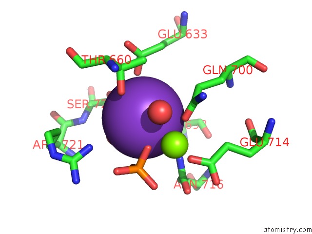
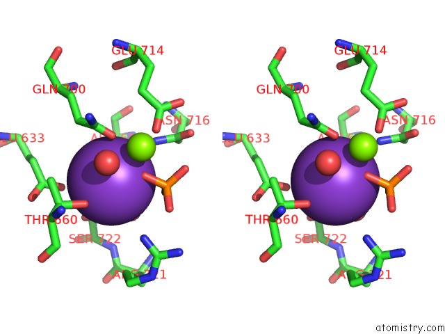
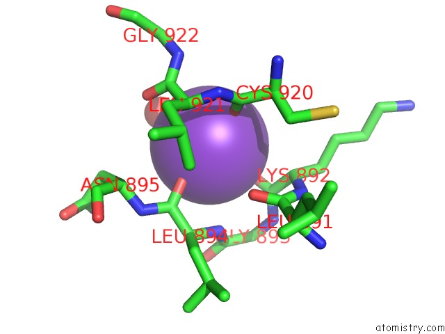
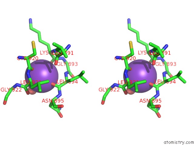
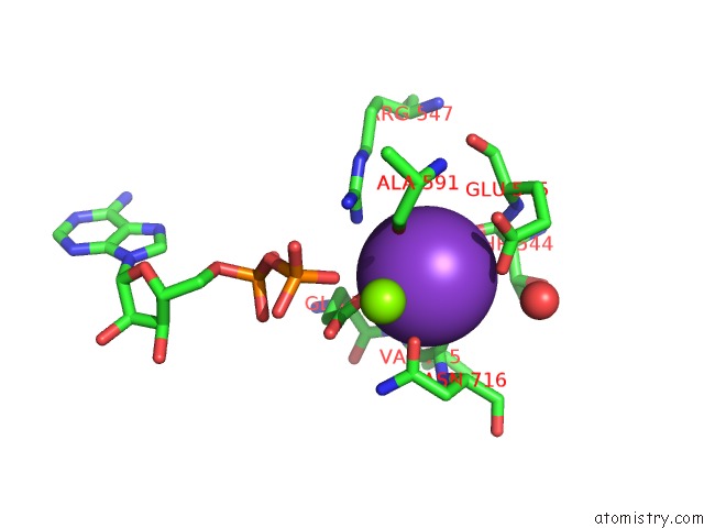
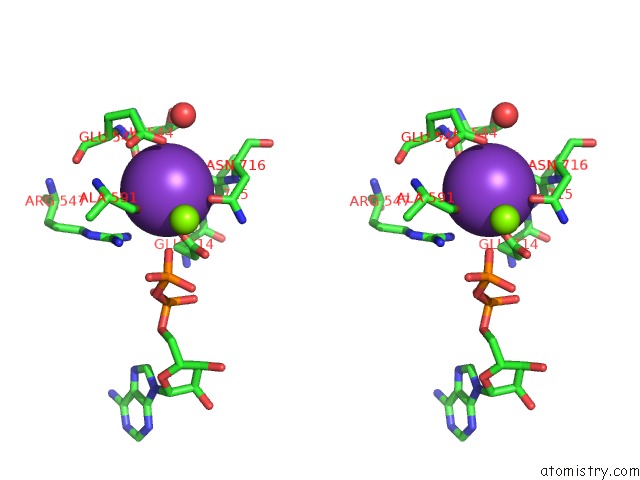
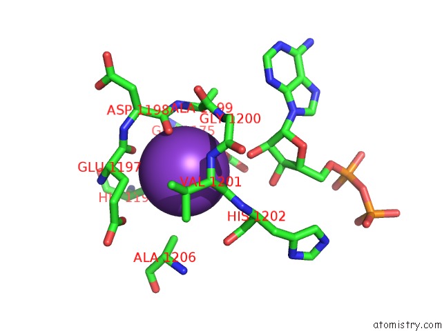
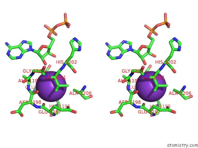
Potassium binding site 1 out of 18 in 5dou
Go back to
Potassium binding site 1 out
of 18 in the Crystal Structure of Human Carbamoyl Phosphate Synthetase I (CPS1), Ligand-Bound Form
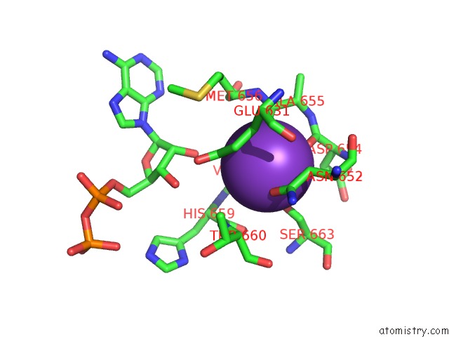
Mono view
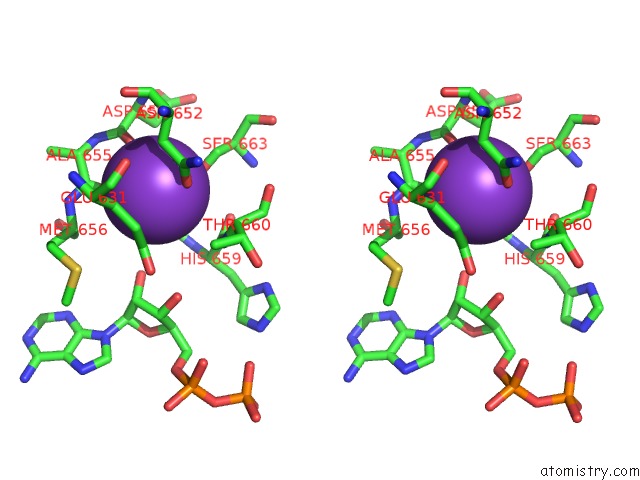
Stereo pair view
Mono view
Stereo pair view
A full contact list of Potassium with other atoms in the K binding
site number 1 of Crystal Structure of Human Carbamoyl Phosphate Synthetase I (CPS1), Ligand-Bound Form within 5.0Å range:
|
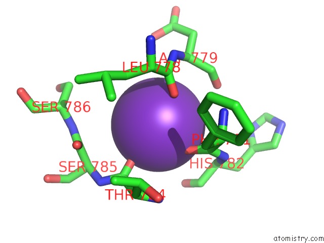
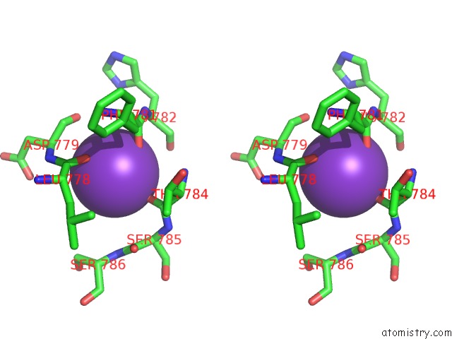
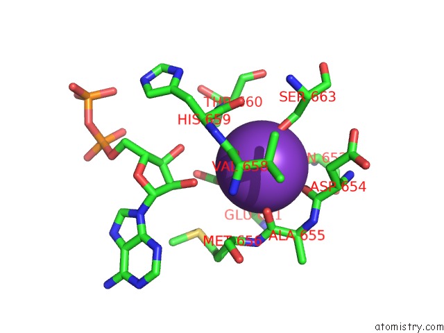
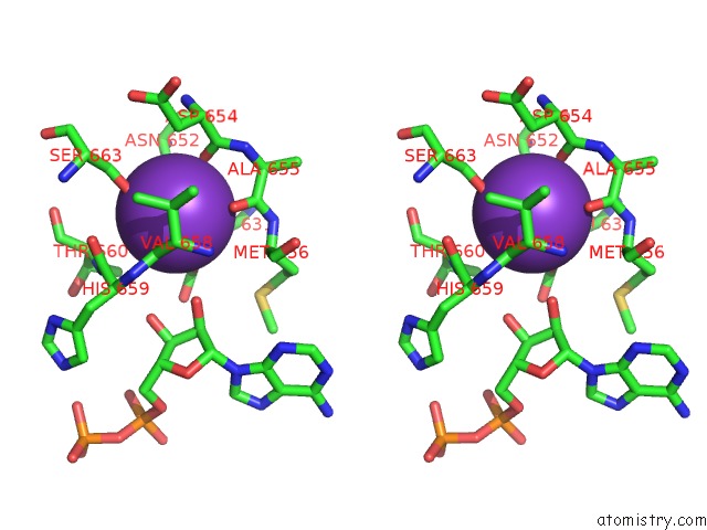
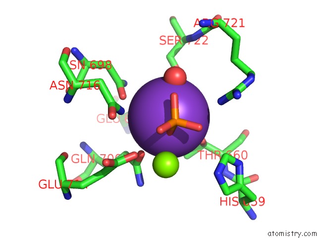
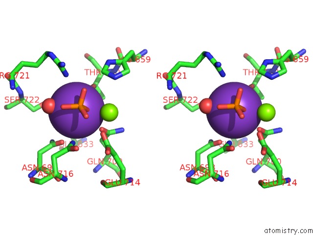
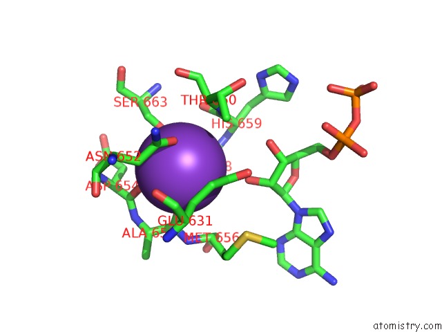
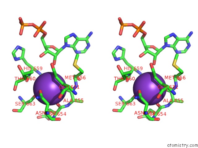
Potassium binding site 2 out of 18 in 5dou
Go back to
Potassium binding site 2 out
of 18 in the Crystal Structure of Human Carbamoyl Phosphate Synthetase I (CPS1), Ligand-Bound Form
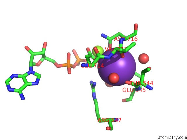
Mono view
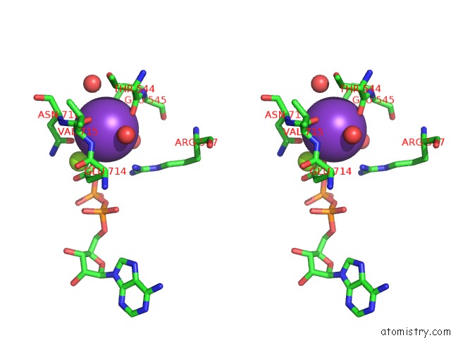
Stereo pair view
Mono view
Stereo pair view
A full contact list of Potassium with other atoms in the K binding
site number 2 of Crystal Structure of Human Carbamoyl Phosphate Synthetase I (CPS1), Ligand-Bound Form within 5.0Å range:
|
Potassium binding site 3 out of 18 in 5dou
Go back to
Potassium binding site 3 out
of 18 in the Crystal Structure of Human Carbamoyl Phosphate Synthetase I (CPS1), Ligand-Bound Form
Mono view
Stereo pair view
Mono view
Stereo pair view
A full contact list of Potassium with other atoms in the K binding
site number 3 of Crystal Structure of Human Carbamoyl Phosphate Synthetase I (CPS1), Ligand-Bound Form within 5.0Å range:
|
Potassium binding site 4 out of 18 in 5dou
Go back to
Potassium binding site 4 out
of 18 in the Crystal Structure of Human Carbamoyl Phosphate Synthetase I (CPS1), Ligand-Bound Form
Mono view
Stereo pair view
Mono view
Stereo pair view
A full contact list of Potassium with other atoms in the K binding
site number 4 of Crystal Structure of Human Carbamoyl Phosphate Synthetase I (CPS1), Ligand-Bound Form within 5.0Å range:
|
Potassium binding site 5 out of 18 in 5dou
Go back to
Potassium binding site 5 out
of 18 in the Crystal Structure of Human Carbamoyl Phosphate Synthetase I (CPS1), Ligand-Bound Form
Mono view
Stereo pair view
Mono view
Stereo pair view
A full contact list of Potassium with other atoms in the K binding
site number 5 of Crystal Structure of Human Carbamoyl Phosphate Synthetase I (CPS1), Ligand-Bound Form within 5.0Å range:
|
Potassium binding site 6 out of 18 in 5dou
Go back to
Potassium binding site 6 out
of 18 in the Crystal Structure of Human Carbamoyl Phosphate Synthetase I (CPS1), Ligand-Bound Form
Mono view
Stereo pair view
Mono view
Stereo pair view
A full contact list of Potassium with other atoms in the K binding
site number 6 of Crystal Structure of Human Carbamoyl Phosphate Synthetase I (CPS1), Ligand-Bound Form within 5.0Å range:
|
Potassium binding site 7 out of 18 in 5dou
Go back to
Potassium binding site 7 out
of 18 in the Crystal Structure of Human Carbamoyl Phosphate Synthetase I (CPS1), Ligand-Bound Form
Mono view
Stereo pair view
Mono view
Stereo pair view
A full contact list of Potassium with other atoms in the K binding
site number 7 of Crystal Structure of Human Carbamoyl Phosphate Synthetase I (CPS1), Ligand-Bound Form within 5.0Å range:
|
Potassium binding site 8 out of 18 in 5dou
Go back to
Potassium binding site 8 out
of 18 in the Crystal Structure of Human Carbamoyl Phosphate Synthetase I (CPS1), Ligand-Bound Form
Mono view
Stereo pair view
Mono view
Stereo pair view
A full contact list of Potassium with other atoms in the K binding
site number 8 of Crystal Structure of Human Carbamoyl Phosphate Synthetase I (CPS1), Ligand-Bound Form within 5.0Å range:
|
Potassium binding site 9 out of 18 in 5dou
Go back to
Potassium binding site 9 out
of 18 in the Crystal Structure of Human Carbamoyl Phosphate Synthetase I (CPS1), Ligand-Bound Form
Mono view
Stereo pair view
Mono view
Stereo pair view
A full contact list of Potassium with other atoms in the K binding
site number 9 of Crystal Structure of Human Carbamoyl Phosphate Synthetase I (CPS1), Ligand-Bound Form within 5.0Å range:
|
Potassium binding site 10 out of 18 in 5dou
Go back to
Potassium binding site 10 out
of 18 in the Crystal Structure of Human Carbamoyl Phosphate Synthetase I (CPS1), Ligand-Bound Form
Mono view
Stereo pair view
Mono view
Stereo pair view
A full contact list of Potassium with other atoms in the K binding
site number 10 of Crystal Structure of Human Carbamoyl Phosphate Synthetase I (CPS1), Ligand-Bound Form within 5.0Å range:
|
Reference:
S.De Cima,
L.M.Polo,
C.Diez-Fernandez,
A.I.Martinez,
J.Cervera,
I.Fita,
V.Rubio.
Structure of Human Carbamoyl Phosphate Synthetase: Deciphering the on/Off Switch of Human Ureagenesis. Sci Rep V. 5 16950 2015.
ISSN: ESSN 2045-2322
PubMed: 26592762
DOI: 10.1038/SREP16950
Page generated: Mon Aug 12 13:17:18 2024
ISSN: ESSN 2045-2322
PubMed: 26592762
DOI: 10.1038/SREP16950
Last articles
Zn in 9J0NZn in 9J0O
Zn in 9J0P
Zn in 9FJX
Zn in 9EKB
Zn in 9C0F
Zn in 9CAH
Zn in 9CH0
Zn in 9CH3
Zn in 9CH1