Potassium »
PDB 5cbv-5dea »
5dav »
Potassium in PDB 5dav: Fe(II)/(Alpha)Ketoglutarate-Dependent Dioxygenase Asqj in Complex with 4-Methoxydehydrocyclopeptin
Protein crystallography data
The structure of Fe(II)/(Alpha)Ketoglutarate-Dependent Dioxygenase Asqj in Complex with 4-Methoxydehydrocyclopeptin, PDB code: 5dav
was solved by
M.Groll,
A.Braeuer,
with X-Ray Crystallography technique. A brief refinement statistics is given in the table below:
| Resolution Low / High (Å) | 15.00 / 1.80 |
| Space group | C 2 2 21 |
| Cell size a, b, c (Å), α, β, γ (°) | 73.050, 120.770, 66.660, 90.00, 90.00, 90.00 |
| R / Rfree (%) | 16.4 / 19.6 |
Other elements in 5dav:
The structure of Fe(II)/(Alpha)Ketoglutarate-Dependent Dioxygenase Asqj in Complex with 4-Methoxydehydrocyclopeptin also contains other interesting chemical elements:
| Nickel | (Ni) | 1 atom |
| Bromine | (Br) | 2 atoms |
Potassium Binding Sites:
The binding sites of Potassium atom in the Fe(II)/(Alpha)Ketoglutarate-Dependent Dioxygenase Asqj in Complex with 4-Methoxydehydrocyclopeptin
(pdb code 5dav). This binding sites where shown within
5.0 Angstroms radius around Potassium atom.
In total only one binding site of Potassium was determined in the Fe(II)/(Alpha)Ketoglutarate-Dependent Dioxygenase Asqj in Complex with 4-Methoxydehydrocyclopeptin, PDB code: 5dav:
In total only one binding site of Potassium was determined in the Fe(II)/(Alpha)Ketoglutarate-Dependent Dioxygenase Asqj in Complex with 4-Methoxydehydrocyclopeptin, PDB code: 5dav:
Potassium binding site 1 out of 1 in 5dav
Go back to
Potassium binding site 1 out
of 1 in the Fe(II)/(Alpha)Ketoglutarate-Dependent Dioxygenase Asqj in Complex with 4-Methoxydehydrocyclopeptin
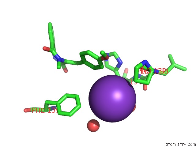
Mono view
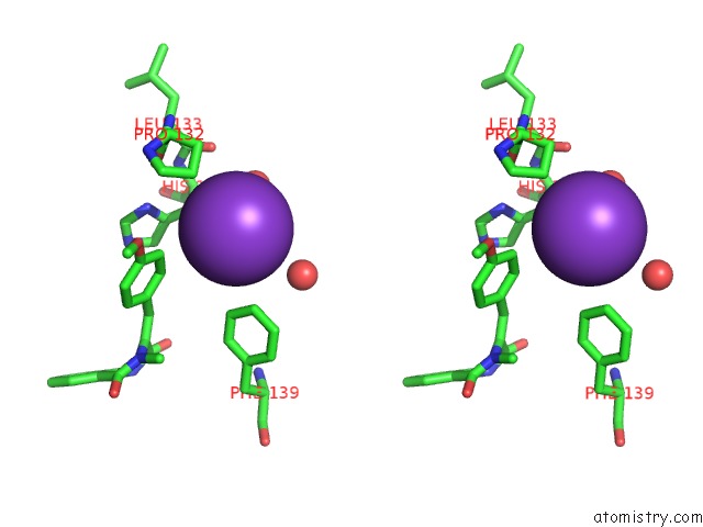
Stereo pair view
Mono view
Stereo pair view
A full contact list of Potassium with other atoms in the K binding
site number 1 of Fe(II)/(Alpha)Ketoglutarate-Dependent Dioxygenase Asqj in Complex with 4-Methoxydehydrocyclopeptin within 5.0Å range:
|
Reference:
A.Brauer,
P.Beck,
L.Hintermann,
M.Groll.
Structure of the Dioxygenase Asqj: Mechanistic Insights Into A One-Pot Multistep Quinolone Antibiotic Biosynthesis. Angew.Chem.Int.Ed.Engl. V. 55 422 2016.
ISSN: ESSN 1521-3773
PubMed: 26553478
DOI: 10.1002/ANIE.201507835
Page generated: Mon Aug 12 13:12:20 2024
ISSN: ESSN 1521-3773
PubMed: 26553478
DOI: 10.1002/ANIE.201507835
Last articles
Zn in 9JYWZn in 9IR4
Zn in 9IR3
Zn in 9GMX
Zn in 9GMW
Zn in 9JEJ
Zn in 9ERF
Zn in 9ERE
Zn in 9EGV
Zn in 9EGW