Potassium »
PDB 5cbv-5dea »
5cmx »
Potassium in PDB 5cmx: X-Ray Structure of the Complex Between Human Alpha Thrombin and A Duplex/Quadruplex 31-Mer Dna Aptamer
Enzymatic activity of X-Ray Structure of the Complex Between Human Alpha Thrombin and A Duplex/Quadruplex 31-Mer Dna Aptamer
All present enzymatic activity of X-Ray Structure of the Complex Between Human Alpha Thrombin and A Duplex/Quadruplex 31-Mer Dna Aptamer:
3.4.21.5;
3.4.21.5;
Protein crystallography data
The structure of X-Ray Structure of the Complex Between Human Alpha Thrombin and A Duplex/Quadruplex 31-Mer Dna Aptamer, PDB code: 5cmx
was solved by
I.Russo Krauss,
A.Pica,
V.Napolitano,
F.Sica,
with X-Ray Crystallography technique. A brief refinement statistics is given in the table below:
| Resolution Low / High (Å) | 65.67 / 2.98 |
| Space group | P 21 21 2 |
| Cell size a, b, c (Å), α, β, γ (°) | 77.319, 124.420, 40.440, 90.00, 90.00, 90.00 |
| R / Rfree (%) | 16.6 / 23.1 |
Potassium Binding Sites:
The binding sites of Potassium atom in the X-Ray Structure of the Complex Between Human Alpha Thrombin and A Duplex/Quadruplex 31-Mer Dna Aptamer
(pdb code 5cmx). This binding sites where shown within
5.0 Angstroms radius around Potassium atom.
In total only one binding site of Potassium was determined in the X-Ray Structure of the Complex Between Human Alpha Thrombin and A Duplex/Quadruplex 31-Mer Dna Aptamer, PDB code: 5cmx:
In total only one binding site of Potassium was determined in the X-Ray Structure of the Complex Between Human Alpha Thrombin and A Duplex/Quadruplex 31-Mer Dna Aptamer, PDB code: 5cmx:
Potassium binding site 1 out of 1 in 5cmx
Go back to
Potassium binding site 1 out
of 1 in the X-Ray Structure of the Complex Between Human Alpha Thrombin and A Duplex/Quadruplex 31-Mer Dna Aptamer
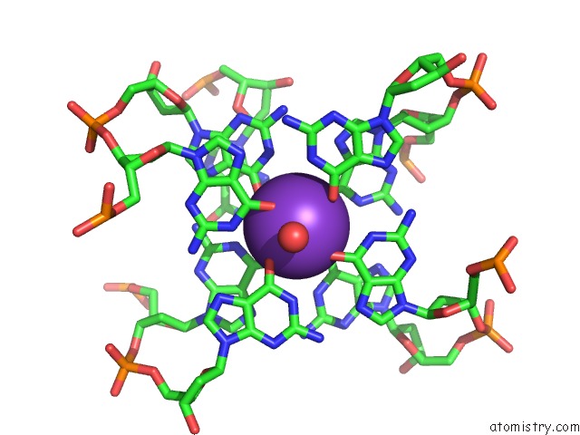
Mono view
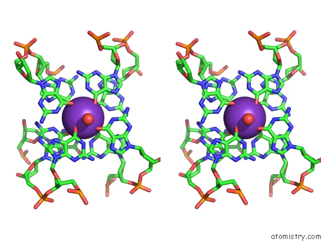
Stereo pair view
Mono view
Stereo pair view
A full contact list of Potassium with other atoms in the K binding
site number 1 of X-Ray Structure of the Complex Between Human Alpha Thrombin and A Duplex/Quadruplex 31-Mer Dna Aptamer within 5.0Å range:
|
Reference:
I.Russo Krauss,
V.Spiridonova,
A.Pica,
V.Napolitano,
F.Sica.
Different Duplex/Quadruplex Junctions Determine the Properties of Anti-Thrombin Aptamers with Mixed Folding. Nucleic Acids Res. V. 44 983 2016.
ISSN: ESSN 1362-4962
PubMed: 26673709
DOI: 10.1093/NAR/GKV1384
Page generated: Mon Aug 12 13:08:40 2024
ISSN: ESSN 1362-4962
PubMed: 26673709
DOI: 10.1093/NAR/GKV1384
Last articles
Zn in 9J0NZn in 9J0O
Zn in 9J0P
Zn in 9FJX
Zn in 9EKB
Zn in 9C0F
Zn in 9CAH
Zn in 9CH0
Zn in 9CH3
Zn in 9CH1