Potassium »
PDB 5avx-5cbu »
5c8c »
Potassium in PDB 5c8c: Crystal Structure of Recombinant Coxsackievirus A16 Capsid
Protein crystallography data
The structure of Crystal Structure of Recombinant Coxsackievirus A16 Capsid, PDB code: 5c8c
was solved by
J.Ren,
X.Wang,
L.Zhu,
Z.Hu,
Q.Gao,
P.Yang,
X.Li,
J.Wang,
X.Shen,
E.E.Fry,
Z.Rao,
D.I.Stuart,
with X-Ray Crystallography technique. A brief refinement statistics is given in the table below:
| Resolution Low / High (Å) | 49.70 / 2.50 |
| Space group | P 42 3 2 1 |
| Cell size a, b, c (Å), α, β, γ (°) | 347.900, 347.900, 347.900, 90.00, 90.00, 90.00 |
| R / Rfree (%) | 16.4 / 17.2 |
Other elements in 5c8c:
The structure of Crystal Structure of Recombinant Coxsackievirus A16 Capsid also contains other interesting chemical elements:
| Chlorine | (Cl) | 2 atoms |
Potassium Binding Sites:
The binding sites of Potassium atom in the Crystal Structure of Recombinant Coxsackievirus A16 Capsid
(pdb code 5c8c). This binding sites where shown within
5.0 Angstroms radius around Potassium atom.
In total only one binding site of Potassium was determined in the Crystal Structure of Recombinant Coxsackievirus A16 Capsid, PDB code: 5c8c:
In total only one binding site of Potassium was determined in the Crystal Structure of Recombinant Coxsackievirus A16 Capsid, PDB code: 5c8c:
Potassium binding site 1 out of 1 in 5c8c
Go back to
Potassium binding site 1 out
of 1 in the Crystal Structure of Recombinant Coxsackievirus A16 Capsid
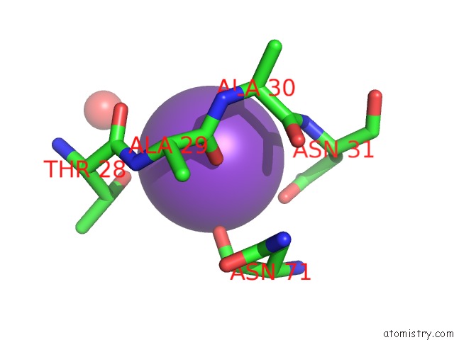
Mono view
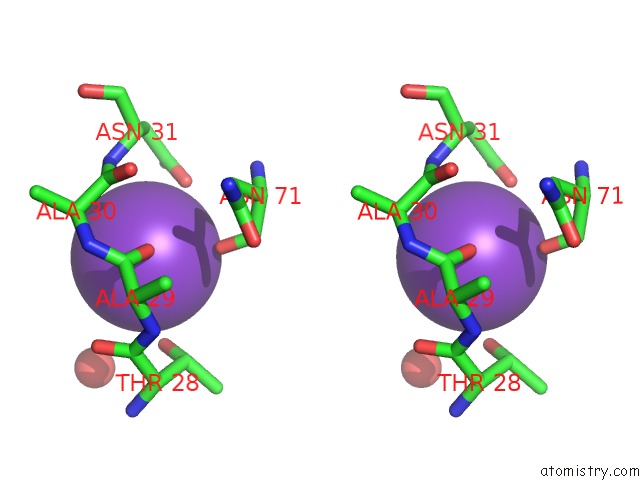
Stereo pair view
Mono view
Stereo pair view
A full contact list of Potassium with other atoms in the K binding
site number 1 of Crystal Structure of Recombinant Coxsackievirus A16 Capsid within 5.0Å range:
|
Reference:
J.Ren,
X.Wang,
L.Zhu,
Z.Hu,
Q.Gao,
P.Yang,
X.Li,
J.Wang,
X.Shen,
E.E.Fry,
Z.Rao,
D.I.Stuart.
Structures of Coxsackievirus A16 Capsids with Native Antigenicity: Implications For Particle Expansion, Receptor Binding, and Immunogenicity. J.Virol. V. 89 10500 2015.
ISSN: ESSN 1098-5514
PubMed: 26269176
DOI: 10.1128/JVI.01102-15
Page generated: Mon Aug 12 13:05:25 2024
ISSN: ESSN 1098-5514
PubMed: 26269176
DOI: 10.1128/JVI.01102-15
Last articles
Zn in 9MJ5Zn in 9HNW
Zn in 9G0L
Zn in 9FNE
Zn in 9DZN
Zn in 9E0I
Zn in 9D32
Zn in 9DAK
Zn in 8ZXC
Zn in 8ZUF