Potassium »
PDB 4zun-5avw »
5aop »
Potassium in PDB 5aop: Sulfite Reductase Structure Reduced with Crii Edta, 5-Coordinate Siroheme, Siroheme Feii, [4FE-4S] +1
Enzymatic activity of Sulfite Reductase Structure Reduced with Crii Edta, 5-Coordinate Siroheme, Siroheme Feii, [4FE-4S] +1
All present enzymatic activity of Sulfite Reductase Structure Reduced with Crii Edta, 5-Coordinate Siroheme, Siroheme Feii, [4FE-4S] +1:
1.8.1.2;
1.8.1.2;
Protein crystallography data
The structure of Sulfite Reductase Structure Reduced with Crii Edta, 5-Coordinate Siroheme, Siroheme Feii, [4FE-4S] +1, PDB code: 5aop
was solved by
B.R.Crane,
E.D.Getzoff,
with X-Ray Crystallography technique. A brief refinement statistics is given in the table below:
| Resolution Low / High (Å) | 10.00 / 2.20 |
| Space group | P 21 21 21 |
| Cell size a, b, c (Å), α, β, γ (°) | 69.800, 77.400, 87.800, 90.00, 90.00, 90.00 |
| R / Rfree (%) | 16.4 / n/a |
Other elements in 5aop:
The structure of Sulfite Reductase Structure Reduced with Crii Edta, 5-Coordinate Siroheme, Siroheme Feii, [4FE-4S] +1 also contains other interesting chemical elements:
| Iron | (Fe) | 5 atoms |
Potassium Binding Sites:
The binding sites of Potassium atom in the Sulfite Reductase Structure Reduced with Crii Edta, 5-Coordinate Siroheme, Siroheme Feii, [4FE-4S] +1
(pdb code 5aop). This binding sites where shown within
5.0 Angstroms radius around Potassium atom.
In total only one binding site of Potassium was determined in the Sulfite Reductase Structure Reduced with Crii Edta, 5-Coordinate Siroheme, Siroheme Feii, [4FE-4S] +1, PDB code: 5aop:
In total only one binding site of Potassium was determined in the Sulfite Reductase Structure Reduced with Crii Edta, 5-Coordinate Siroheme, Siroheme Feii, [4FE-4S] +1, PDB code: 5aop:
Potassium binding site 1 out of 1 in 5aop
Go back to
Potassium binding site 1 out
of 1 in the Sulfite Reductase Structure Reduced with Crii Edta, 5-Coordinate Siroheme, Siroheme Feii, [4FE-4S] +1
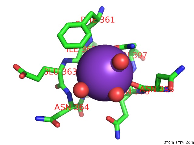
Mono view
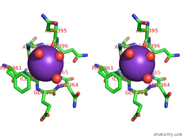
Stereo pair view
Mono view
Stereo pair view
A full contact list of Potassium with other atoms in the K binding
site number 1 of Sulfite Reductase Structure Reduced with Crii Edta, 5-Coordinate Siroheme, Siroheme Feii, [4FE-4S] +1 within 5.0Å range:
|
Reference:
B.R.Crane,
L.M.Siegel,
E.D.Getzoff.
Structures of the Siroheme- and FE4S4-Containing Active Center of Sulfite Reductase in Different States of Oxidation: Heme Activation Via Reduction-Gated Exogenous Ligand Exchange. Biochemistry V. 36 12101 1997.
ISSN: ISSN 0006-2960
PubMed: 9315848
DOI: 10.1021/BI971065Q
Page generated: Mon Aug 12 12:55:29 2024
ISSN: ISSN 0006-2960
PubMed: 9315848
DOI: 10.1021/BI971065Q
Last articles
Zn in 9J0NZn in 9J0O
Zn in 9J0P
Zn in 9FJX
Zn in 9EKB
Zn in 9C0F
Zn in 9CAH
Zn in 9CH0
Zn in 9CH3
Zn in 9CH1